 Movie
Movie Controller

[English] 日本語
 Yorodumi
- PDB-1mor: ISOMERASE DOMAIN OF GLUCOSAMINE 6-PHOSPHATE SYNTHASE COMPLEXED WI... -
+ Open data

- Basic information
| Entry | Database: PDB / ID: 1mor | ||||||
|---|---|---|---|---|---|---|---|
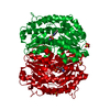
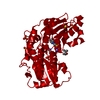
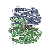
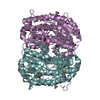
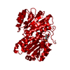
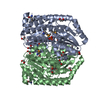
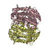
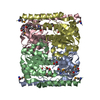
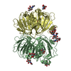
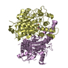
| Title | ISOMERASE DOMAIN OF GLUCOSAMINE 6-PHOSPHATE SYNTHASE COMPLEXED WITH GLUCOSE 6-PHOSPHATE | ||||||
 Components Components | GLUCOSAMINE 6-PHOSPHATE SYNTHASE | ||||||
 Keywords Keywords | GLUTAMINE AMIDOTRANSFERASE | ||||||
| Function / homology |  Function and homology information Function and homology informationglutamine-fructose-6-phosphate transaminase (isomerizing) / L-glutamine:D-fructose-6-phosphate transaminase (isomerizing) activity / UDP-N-acetylglucosamine metabolic process / UDP-N-acetylglucosamine biosynthetic process / carbohydrate derivative binding / fructose 6-phosphate metabolic process / protein N-linked glycosylation / carbohydrate metabolic process / cytosol Similarity search - Function | ||||||
| Biological species |  | ||||||
| Method |  X-RAY DIFFRACTION / SYNCHROTRON / MIR / Resolution: 1.9 Å X-RAY DIFFRACTION / SYNCHROTRON / MIR / Resolution: 1.9 Å | ||||||
 Authors Authors | Teplyakov, A. | ||||||
 Citation Citation | Journal: Protein Sci. / Year: 1999 Title: The mechanism of sugar phosphate isomerization by glucosamine 6-phosphate synthase. Authors: Teplyakov, A. / Obmolova, G. / Badet-Denisot, M.A. / Badet, B. #1: Journal: Protein Sci. / Year: 1999Title: The Mechanism of Sugar Phosphate Isomerization by Glucosamine 6-Phosphate Synthase Authors: Teplyakov, A. / Obmolova, G. / Badet-Denisot, M.A. / Badet, B. #2: Journal: Structure / Year: 1997Title: Erratum. Substrate Binding is Required for Assembly of the Active Conformation of the Catalytic Site in Ntn Amidotransferases: Evidence from the 1.8 A Crystal Structure of the Glutaminase ...Title: Erratum. Substrate Binding is Required for Assembly of the Active Conformation of the Catalytic Site in Ntn Amidotransferases: Evidence from the 1.8 A Crystal Structure of the Glutaminase Domain of Glucosamine 6-Phosphate Synthase Authors: Isupov, M.N. / Obmolova, G. / Butterworth, S. / Badet-Denisot, M.A. / Badet, B. / Polikarpov, I. / Littlechild, J.A. / Teplyakov, A. #3: Journal: Structure / Year: 1996Title: Substrate Binding is Required for Assembly of the Active Conformation of the Catalytic Site in Ntn Amidotransferases: Evidence from the 1.8 A Crystal Structure of the Glutaminase Domain of ...Title: Substrate Binding is Required for Assembly of the Active Conformation of the Catalytic Site in Ntn Amidotransferases: Evidence from the 1.8 A Crystal Structure of the Glutaminase Domain of Glucosamine 6-Phosphate Synthase Authors: Isupov, M.N. / Obmolova, G. / Butterworth, S. / Badet-Denisot, M.A. / Badet, B. / Polikarpov, I. / Littlechild, J.A. / Teplyakov, A. #4: Journal: J.Mol.Biol. / Year: 1994Title: Crystallization and Preliminary X-Ray Analysis of the Two Domains of Glucosamine-6-Phosphate Synthase from Escherichia Coli Authors: Obmolova, G. / Badet-Denisot, M.A. / Badet, B. / Teplyakov, A. | ||||||
| History |
|
- Structure visualization
| Structure viewer | Molecule: MolmilJmol/JSmol |
|---|
- Downloads & links
-Download
| PDBx/mmCIF format | 1mor.cif.gz | 87.4 KB | Display | PDBx/mmCIF format |
|---|---|---|---|---|
| PDB format | pdb1mor.ent.gz | 66.3 KB | Display | PDB format |
| PDBx/mmJSON format | 1mor.json.gz | Tree view | PDBx/mmJSON format | |
| Others |  Other downloads Other downloads |
-Validation report
| Arichive directory | https://data.pdbj.org/pub/pdb/validation_reports/mo/1morftp://data.pdbj.org/pub/pdb/validation_reports/mo/1mor | HTTPS FTP |
|---|
-Related structure data

-Links
 PDBj
PDBj


- Assembly
| Deposited unit | 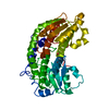
| ||||||||
|---|---|---|---|---|---|---|---|---|---|
| 1 | 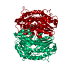
| ||||||||
| 2 | x 6
| ||||||||
| Unit cell |
|
-Components
| #1: Protein | Mass: 40357.004 Da / Num. of mol.: 1 Source method: isolated from a genetically manipulated source Source: (gene. exp.) References: UniProt: P17169, glutamine-fructose-6-phosphate transaminase (isomerizing) |
|---|---|
| #2: Sugar | ChemComp-G6P /   Type: D-saccharide, alpha linking / Mass: 260.136 Da / Num. of mol.: 1 Type: D-saccharide, alpha linking / Mass: 260.136 Da / Num. of mol.: 1Source method: isolated from a genetically manipulated source Formula: C6H13O9P |
| #3: Water | ChemComp-HOH /  Mass: 18.015 Da / Num. of mol.: 223 / Source method: isolated from a natural source / Formula: H2O Mass: 18.015 Da / Num. of mol.: 223 / Source method: isolated from a natural source / Formula: H2O |
-Experimental details
-Experiment
| Experiment | Method: X-RAY DIFFRACTION / Number of used crystals: 2 |
|---|
- Sample preparation
| Crystal | Density Matthews: 4.4 Å3/Da / Density % sol: 72 % | |||||||||||||||||||||||||
|---|---|---|---|---|---|---|---|---|---|---|---|---|---|---|---|---|---|---|---|---|---|---|---|---|---|---|
| Crystal grow | pH: 6 / Details: pH 6.0 | |||||||||||||||||||||||||
| Crystal | *PLUS Density % sol: 72 % | |||||||||||||||||||||||||
| Crystal grow | *PLUS Method: unknown | |||||||||||||||||||||||||
| Components of the solutions | *PLUS
|
-Data collection
| Diffraction | Mean temperature: 277 K |
|---|---|
| Diffraction source | Source: SYNCHROTRON / Site: EMBL/DESY, HAMBURG  / Beamline: X11 / Wavelength: 0.912 / Beamline: X11 / Wavelength: 0.912 |
| Detector | Type: MAR scanner 300 mm plate / Detector: IMAGE PLATE / Date: Aug 28, 1996 / Details: CYLINDRICAL MIRROR |
| Radiation | Monochromator: GE(111) / Monochromatic (M) / Laue (L): M / Scattering type: x-ray |
| Radiation wavelength | Wavelength: 0.912 Å / Relative weight: 1 |
| Reflection | Resolution: 1.9→20 Å / Num. obs: 205104 / % possible obs: 99.2 % / Observed criterion σ(I): -3 / Redundancy: 3.7 % / Biso Wilson estimate: 26 Å2 / Rmerge(I) obs: 0.084 / Net I/σ(I): 14.3 |
| Reflection shell | Resolution: 1.9→1.93 Å / Redundancy: 2.9 % / Rmerge(I) obs: 0.593 / Mean I/σ(I) obs: 1.8 / % possible all: 98.8 |
| Reflection | *PLUS Num. obs: 55613 |
- Processing
| Software |
| ||||||||||||||||||||||||||||||||||||||||||||||||||||||||||||||||||||||||||||||||||||
|---|---|---|---|---|---|---|---|---|---|---|---|---|---|---|---|---|---|---|---|---|---|---|---|---|---|---|---|---|---|---|---|---|---|---|---|---|---|---|---|---|---|---|---|---|---|---|---|---|---|---|---|---|---|---|---|---|---|---|---|---|---|---|---|---|---|---|---|---|---|---|---|---|---|---|---|---|---|---|---|---|---|---|---|---|---|
| Refinement | Method to determine structure: MIR / Resolution: 1.9→10 Å / σ(F): 0
| ||||||||||||||||||||||||||||||||||||||||||||||||||||||||||||||||||||||||||||||||||||
| Displacement parameters | Biso mean: 31.1 Å2 | ||||||||||||||||||||||||||||||||||||||||||||||||||||||||||||||||||||||||||||||||||||
| Refine analyze | Luzzati coordinate error obs: 0.25 Å | ||||||||||||||||||||||||||||||||||||||||||||||||||||||||||||||||||||||||||||||||||||
| Refinement step | Cycle: LAST / Resolution: 1.9→10 Å
| ||||||||||||||||||||||||||||||||||||||||||||||||||||||||||||||||||||||||||||||||||||
| Refine LS restraints |
| ||||||||||||||||||||||||||||||||||||||||||||||||||||||||||||||||||||||||||||||||||||
| Software | *PLUS Name: PROLSQ / Classification: refinement | ||||||||||||||||||||||||||||||||||||||||||||||||||||||||||||||||||||||||||||||||||||
| Refinement | *PLUS % reflection Rfree: 5 % / Rfactor obs: 0.191 / Rfactor Rfree: 0.224 | ||||||||||||||||||||||||||||||||||||||||||||||||||||||||||||||||||||||||||||||||||||
| Solvent computation | *PLUS | ||||||||||||||||||||||||||||||||||||||||||||||||||||||||||||||||||||||||||||||||||||
| Displacement parameters | *PLUS |
