 Movie
Movie Controller

[English] 日本語
 Yorodumi
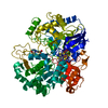
- PDB-2zj3: Isomerase domain of human glucose:fructose-6-phosphate amidotrans... -
+ Open data

- Basic information
| Entry | Database: PDB / ID: 2zj3 | ||||||
|---|---|---|---|---|---|---|---|
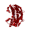
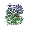
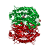
| Title | Isomerase domain of human glucose:fructose-6-phosphate amidotransferase | ||||||
 Components Components | Glucosamine--fructose-6-phosphate aminotransferase [isomerizing] 1 | ||||||
 Keywords Keywords | TRANSFERASE / glucosamine-6-phosphate synthase / aldose/ketose isomerase / Rossmann-like fold / Alternative splicing / Aminotransferase / Glutamine amidotransferase / Phosphoprotein | ||||||
| Function / homology |  Function and homology information Function and homology informationDefective GFPT1 causes CMSTA1 / Synthesis of UDP-N-acetyl-glucosamine / glutamine-fructose-6-phosphate transaminase (isomerizing) / L-glutamine:D-fructose-6-phosphate transaminase (isomerizing) activity / UDP-N-acetylglucosamine metabolic process / UDP-N-acetylglucosamine biosynthetic process / XBP1(S) activates chaperone genes / energy reserve metabolic process / carbohydrate derivative binding / fructose 6-phosphate metabolic process ...Defective GFPT1 causes CMSTA1 / Synthesis of UDP-N-acetyl-glucosamine / glutamine-fructose-6-phosphate transaminase (isomerizing) / L-glutamine:D-fructose-6-phosphate transaminase (isomerizing) activity / UDP-N-acetylglucosamine metabolic process / UDP-N-acetylglucosamine biosynthetic process / XBP1(S) activates chaperone genes / energy reserve metabolic process / carbohydrate derivative binding / fructose 6-phosphate metabolic process / protein N-linked glycosylation / circadian regulation of gene expression / extracellular exosome / cytosol Similarity search - Function | ||||||
| Biological species |  Homo sapiens (human) Homo sapiens (human) | ||||||
| Method |  X-RAY DIFFRACTION / SYNCHROTRON / MOLECULAR REPLACEMENT / Resolution: 1.9 Å X-RAY DIFFRACTION / SYNCHROTRON / MOLECULAR REPLACEMENT / Resolution: 1.9 Å | ||||||
 Authors Authors | Nakaishi, Y. / Bando, M. / Kondo, K. / Tsuge, H. | ||||||
 Citation Citation | Journal: Febs Lett. / Year: 2009 Title: Structural analysis of human glutamine:fructose-6-phosphate amidotransferase, a key regulator in type 2 diabetes Authors: Nakaishi, Y. / Bando, M. / Shimizu, H. / Watanabe, K. / Goto, F. / Tsuge, H. / Kondo, K. / Komatsu, M. | ||||||
| History |
|
- Structure visualization
| Structure viewer | Molecule: MolmilJmol/JSmol |
|---|
- Downloads & links
-Download
| PDBx/mmCIF format | 2zj3.cif.gz | 91.3 KB | Display | PDBx/mmCIF format |
|---|---|---|---|---|
| PDB format | pdb2zj3.ent.gz | 68.7 KB | Display | PDB format |
| PDBx/mmJSON format | 2zj3.json.gz | Tree view | PDBx/mmJSON format | |
| Others |  Other downloads Other downloads |
-Validation report
| Arichive directory | https://data.pdbj.org/pub/pdb/validation_reports/zj/2zj3ftp://data.pdbj.org/pub/pdb/validation_reports/zj/2zj3 | HTTPS FTP |
|---|
-Related structure data
| Related structure data |  2zj4C 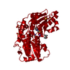 1moqS C: citing same article ( S: Starting model for refinement |
|---|---|
| Similar structure data |

-Links
 PDBj
PDBj


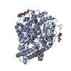
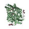
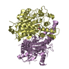
- Assembly
| Deposited unit | 
| ||||||||
|---|---|---|---|---|---|---|---|---|---|
| 1 | 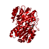
| ||||||||
| Unit cell |
| ||||||||
| Components on special symmetry positions |
|
-Components
| #1: Protein | Mass: 42319.133 Da / Num. of mol.: 1 Fragment: SIS(Sugar ISomerase) domains, UNP Residues 332-699 Source method: isolated from a genetically manipulated source Source: (gene. exp.) Homo sapiens (human) / Plasmid: pET23 / Production host:  References: UniProt: Q06210, glutamine-fructose-6-phosphate transaminase (isomerizing) |
|---|---|
| #2: Sugar | ChemComp-G6P / 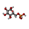  Type: D-saccharide, alpha linking / Mass: 260.136 Da / Num. of mol.: 1 Type: D-saccharide, alpha linking / Mass: 260.136 Da / Num. of mol.: 1Source method: isolated from a genetically manipulated source Formula: C6H13O9P |
| #3: Water | ChemComp-HOH /  Mass: 18.015 Da / Num. of mol.: 277 / Source method: isolated from a natural source / Formula: H2O Mass: 18.015 Da / Num. of mol.: 277 / Source method: isolated from a natural source / Formula: H2O |
-Experimental details
-Experiment
| Experiment | Method: X-RAY DIFFRACTION / Number of used crystals: 1 |
|---|
- Sample preparation
| Crystal | Density Matthews: 2.51 Å3/Da / Density % sol: 50.95 % |
|---|---|
| Crystal grow | Temperature: 277 K / Method: vapor diffusion, hanging drop / pH: 8.5 Details: 12% Isoporpanol, 0.04M Tris, 0.08M Ammonium acetate, 12% glycerol 10-fold excess of Fructose-6-Phosphate, pH 8.50, VAPOR DIFFUSION, HANGING DROP, temperature 277K |
-Data collection
| Diffraction | Mean temperature: 100 K |
|---|---|
| Diffraction source | Source: SYNCHROTRON / Site: SPring-8  / Beamline: BL32B2 / Wavelength: 1 / Beamline: BL32B2 / Wavelength: 1 |
| Detector | Type: RIGAKU RAXIS V / Detector: IMAGE PLATE / Date: Aug 4, 2003 |
| Radiation | Protocol: SINGLE WAVELENGTH / Monochromatic (M) / Laue (L): M / Scattering type: x-ray |
| Radiation wavelength | Wavelength: 1 Å / Relative weight: 1 |
| Reflection | Resolution: 1.9→29.88 Å / Num. obs: 34205 / % possible obs: 98.9 % |
- Processing
| Software |
| ||||||||||||||||||||||||||||||||||||||||||||||||||||||||||||||||||||||||||||||||||||||||||||||||||||||||||||||||||||||||||||||||||||||||||||||||||||||||||||||||||||||||||
|---|---|---|---|---|---|---|---|---|---|---|---|---|---|---|---|---|---|---|---|---|---|---|---|---|---|---|---|---|---|---|---|---|---|---|---|---|---|---|---|---|---|---|---|---|---|---|---|---|---|---|---|---|---|---|---|---|---|---|---|---|---|---|---|---|---|---|---|---|---|---|---|---|---|---|---|---|---|---|---|---|---|---|---|---|---|---|---|---|---|---|---|---|---|---|---|---|---|---|---|---|---|---|---|---|---|---|---|---|---|---|---|---|---|---|---|---|---|---|---|---|---|---|---|---|---|---|---|---|---|---|---|---|---|---|---|---|---|---|---|---|---|---|---|---|---|---|---|---|---|---|---|---|---|---|---|---|---|---|---|---|---|---|---|---|---|---|---|---|---|---|---|
| Refinement | Method to determine structure: MOLECULAR REPLACEMENT Starting model: PDB ENTRY 1MOQ Resolution: 1.9→29.88 Å / Cor.coef. Fo:Fc: 0.948 / Cor.coef. Fo:Fc free: 0.941 / SU B: 2.696 / SU ML: 0.082 / Cross valid method: THROUGHOUT / ESU R: 0.143 / ESU R Free: 0.131 / Stereochemistry target values: MAXIMUM LIKELIHOOD / Details: HYDROGENS HAVE BEEN ADDED IN THE RIDING POSITIONS
| ||||||||||||||||||||||||||||||||||||||||||||||||||||||||||||||||||||||||||||||||||||||||||||||||||||||||||||||||||||||||||||||||||||||||||||||||||||||||||||||||||||||||||
| Solvent computation | Ion probe radii: 0.8 Å / Shrinkage radii: 0.8 Å / VDW probe radii: 1.4 Å / Solvent model: BABINET MODEL WITH MASK | ||||||||||||||||||||||||||||||||||||||||||||||||||||||||||||||||||||||||||||||||||||||||||||||||||||||||||||||||||||||||||||||||||||||||||||||||||||||||||||||||||||||||||
| Displacement parameters | Biso mean: 20.9 Å2
| ||||||||||||||||||||||||||||||||||||||||||||||||||||||||||||||||||||||||||||||||||||||||||||||||||||||||||||||||||||||||||||||||||||||||||||||||||||||||||||||||||||||||||
| Refinement step | Cycle: LAST / Resolution: 1.9→29.88 Å
| ||||||||||||||||||||||||||||||||||||||||||||||||||||||||||||||||||||||||||||||||||||||||||||||||||||||||||||||||||||||||||||||||||||||||||||||||||||||||||||||||||||||||||
| Refine LS restraints |
| ||||||||||||||||||||||||||||||||||||||||||||||||||||||||||||||||||||||||||||||||||||||||||||||||||||||||||||||||||||||||||||||||||||||||||||||||||||||||||||||||||||||||||
| LS refinement shell | Resolution: 1.9→1.95 Å / Total num. of bins used: 20
|
