 Movie
Movie Controller

[English] 日本語
 Yorodumi
- PDB-3eg5: Crystal structure of MDIA1-TSH GBD-FH3 in complex with CDC42-GMPPNP -
+ Open data

- Basic information
| Entry | Database: PDB / ID: 3eg5 | ||||||
|---|---|---|---|---|---|---|---|
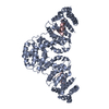
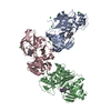
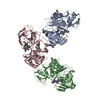
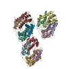
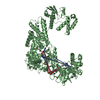
| Title | Crystal structure of MDIA1-TSH GBD-FH3 in complex with CDC42-GMPPNP | ||||||
 Components Components |
| ||||||
 Keywords Keywords | SIGNALING PROTEIN / PROTEIN-PROTEIN COMPLEX / RHO PROTEINS / DIAPHANOUS / FORMINS / ARMADILLO REPEAT / G-PROTEIN / GTPASE / Alternative splicing / Cell membrane / GTP-binding / Lipoprotein / Membrane / Methylation / Nucleotide-binding / Prenylation / Actin-binding / Cell projection / Coiled coil / Cytoplasm / Cytoskeleton / Phosphoprotein / Ubl conjugation | ||||||
| Function / homology |  Function and homology information Function and homology informationDCC mediated attractive signaling / RHO GTPases activate KTN1 / RHOV GTPase cycle / negative regulation of neuron projection regeneration / CD28 dependent Vav1 pathway / multicellular organismal locomotion / RHOQ GTPase cycle / ERBB2 Regulates Cell Motility / RHOF GTPase cycle / actin filament branching ...DCC mediated attractive signaling / RHO GTPases activate KTN1 / RHOV GTPase cycle / negative regulation of neuron projection regeneration / CD28 dependent Vav1 pathway / multicellular organismal locomotion / RHOQ GTPase cycle / ERBB2 Regulates Cell Motility / RHOF GTPase cycle / actin filament branching / RHOU GTPase cycle / RAC3 GTPase cycle / RHOC GTPase cycle / positive regulation of synapse structural plasticity / RHO GTPases activate PAKs / CDC42 GTPase cycle / RAC2 GTPase cycle / nucleus localization / GPVI-mediated activation cascade / : / G beta:gamma signalling through CDC42 / establishment or maintenance of apical/basal cell polarity / GBD domain binding / RHO GTPases Activate WASPs and WAVEs / positive regulation of pinocytosis / COG complex / actin nucleation / neuron projection retraction / EPHB-mediated forward signaling / EGFR downregulation / RAC1 GTPase cycle / RHOB GTPase cycle / cardiac neural crest cell migration involved in outflow tract morphogenesis / storage vacuole / dendritic cell migration / neuron fate determination / RHO GTPases activate IQGAPs / apolipoprotein A-I receptor binding / positive regulation of epithelial cell proliferation involved in lung morphogenesis / endothelin receptor signaling pathway / RHOA GTPase cycle / protein localization to microtubule / regulation of attachment of spindle microtubules to kinetochore / RHOG GTPase cycle / RHO GTPases Activate Formins / organelle transport along microtubule / Regulation of actin dynamics for phagocytic cup formation / Factors involved in megakaryocyte development and platelet production / profilin binding / positive regulation of pseudopodium assembly / MAPK6/MAPK4 signaling / host-mediated perturbation of viral process / positive regulation of intracellular protein transport / regulation of filopodium assembly / cardiac conduction system development / leading edge membrane / heart process / neuropilin signaling pathway / establishment of Golgi localization / Myogenesis / filopodium assembly / cell junction assembly / establishment of epithelial cell apical/basal polarity / dendritic spine morphogenesis / VEGFA-VEGFR2 Pathway / adherens junction organization / GTP-dependent protein binding / thioesterase binding / regulation of lamellipodium assembly / regulation of stress fiber assembly / regulation of modification of postsynaptic structure / embryonic heart tube development / cellular response to histamine / sprouting angiogenesis / regulation of release of sequestered calcium ion into cytosol / regulation of microtubule-based process / axon midline choice point recognition / regulation of postsynapse organization / mitogen-activated protein kinase kinase kinase binding / positive regulation of filopodium assembly / phagocytosis, engulfment / endosomal transport / regulation of mitotic nuclear division / nuclear migration / positive regulation of cytokinesis / spindle midzone / heart contraction / regulation of cytoskeleton organization / establishment of cell polarity / Golgi organization / cell leading edge / establishment or maintenance of cell polarity / lamellipodium membrane / brush border / ephrin receptor signaling pathway / synaptic vesicle endocytosis / negative regulation of protein-containing complex assembly / Rho protein signal transduction / positive regulation of lamellipodium assembly / phagocytic vesicle Similarity search - Function | ||||||
| Biological species |  | ||||||
| Method |  X-RAY DIFFRACTION / SYNCHROTRON / MOLECULAR REPLACEMENT / Resolution: 2.7 Å X-RAY DIFFRACTION / SYNCHROTRON / MOLECULAR REPLACEMENT / Resolution: 2.7 Å | ||||||
 Authors Authors | Lammers, M. / Meyer, S. / Kuehlmann, D. / Wittinghofer, A. | ||||||
 Citation Citation | Journal: J.Biol.Chem. / Year: 2008 Title: Specificity of Interactions between mDia Isoforms and Rho Proteins Authors: Lammers, M. / Meyer, S. / Kuhlmann, D. / Wittinghofer, A. | ||||||
| History |
|
- Structure visualization
| Structure viewer | Molecule: MolmilJmol/JSmol |
|---|
- Downloads & links
-Download
| PDBx/mmCIF format | 3eg5.cif.gz | 214.5 KB | Display | PDBx/mmCIF format |
|---|---|---|---|---|
| PDB format | pdb3eg5.ent.gz | 169.4 KB | Display | PDB format |
| PDBx/mmJSON format | 3eg5.json.gz | Tree view | PDBx/mmJSON format | |
| Others |  Other downloads Other downloads |
-Validation report
| Arichive directory | https://data.pdbj.org/pub/pdb/validation_reports/eg/3eg5ftp://data.pdbj.org/pub/pdb/validation_reports/eg/3eg5 | HTTPS FTP |
|---|
-Related structure data
| Related structure data | 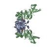 1z2cS S: Starting model for refinement |
|---|---|
| Similar structure data |


-Links
 PDBj
PDBj


- Assembly
| Deposited unit | 
| ||||||||||||||||||||||||||||||||||||||||||||||||||||||||||||||||||||
|---|---|---|---|---|---|---|---|---|---|---|---|---|---|---|---|---|---|---|---|---|---|---|---|---|---|---|---|---|---|---|---|---|---|---|---|---|---|---|---|---|---|---|---|---|---|---|---|---|---|---|---|---|---|---|---|---|---|---|---|---|---|---|---|---|---|---|---|---|---|
| 1 |
| ||||||||||||||||||||||||||||||||||||||||||||||||||||||||||||||||||||
| Unit cell |
| ||||||||||||||||||||||||||||||||||||||||||||||||||||||||||||||||||||
| Noncrystallographic symmetry (NCS) | NCS domain:
NCS domain segments: Component-ID: 1 / Refine code: 1
NCS ensembles :
|
-Components
| #1: Protein | Mass: 19802.719 Da / Num. of mol.: 2 / Fragment: UNP residues 1-178 Source method: isolated from a genetically manipulated source Source: (gene. exp.)  #2: Protein | Mass: 44200.680 Da / Num. of mol.: 2 / Fragment: MDIAN-TSH, UNP residues 69-451 / Mutation: N164T, N165S, N166H Source method: isolated from a genetically manipulated source Source: (gene. exp.) #3: Chemical |   Mass: 522.196 Da / Num. of mol.: 2 / Source method: obtained synthetically / Formula: C10H17N6O13P3 Mass: 522.196 Da / Num. of mol.: 2 / Source method: obtained synthetically / Formula: C10H17N6O13P3Comment: GppNHp, GMPPNP, energy-carrying molecule analogue*YM #4: Chemical |   Mass: 24.305 Da / Num. of mol.: 2 / Source method: obtained synthetically / Formula: Mg Mass: 24.305 Da / Num. of mol.: 2 / Source method: obtained synthetically / Formula: Mg#5: Water | ChemComp-HOH / |  Mass: 18.015 Da / Num. of mol.: 47 / Source method: isolated from a natural source / Formula: H2O Mass: 18.015 Da / Num. of mol.: 47 / Source method: isolated from a natural source / Formula: H2O |
|---|
-Experimental details
-Experiment
| Experiment | Method: X-RAY DIFFRACTION / Number of used crystals: 1 |
|---|
- Sample preparation
| Crystal | Density Matthews: 2.81 Å3/Da / Density % sol: 56.15 % |
|---|---|
| Crystal grow | Temperature: 293 K / Method: vapor diffusion, hanging drop / pH: 8.8 Details: Bis-Tris-Propane (PH 8.8 adjusted with citric acid), 26% (w/v) PEG 3350, 250mM Na-Tartrate, VAPOR DIFFUSION, HANGING DROP, temperature 293K |
-Data collection
| Diffraction | Mean temperature: 100 K |
|---|---|
| Diffraction source | Source: SYNCHROTRON / Site: SLS  / Beamline: X10SA / Wavelength: 0.9763 Å / Beamline: X10SA / Wavelength: 0.9763 Å |
| Detector | Type: MAR CCD 165 mm / Detector: CCD / Date: Apr 25, 2007 / Details: MIRRORS |
| Radiation | Monochromator: SAGITALLY - HORIZONTALLY FOCUSED SI(111) MONOCHROMATOR Protocol: SINGLE WAVELENGTH / Monochromatic (M) / Laue (L): M / Scattering type: x-ray |
| Radiation wavelength | Wavelength: 0.9763 Å / Relative weight: 1 |
| Reflection | Resolution: 2.7→43.478 Å / Num. all: 38241 / Num. obs: 38038 / % possible obs: 99.5 % / Redundancy: 3.2 % / Rmerge(I) obs: 0.074 / Rsym value: 0.084 / Net I/σ(I): 13.52 |
| Reflection shell | Resolution: 2.7→2.8 Å / Redundancy: 3.2 % / Rmerge(I) obs: 0.381 / Mean I/σ(I) obs: 3.69 / Rsym value: 0.333 / % possible all: 99.8 |
- Processing
| Software |
| ||||||||||||||||||||||||||||||||||||||||||||||||||||||||||||||||||||||||||||||||||||||||||||||||||||||||||||||||||||||||||||||||||||||||||||||||||||||||||||||||||||||||||
|---|---|---|---|---|---|---|---|---|---|---|---|---|---|---|---|---|---|---|---|---|---|---|---|---|---|---|---|---|---|---|---|---|---|---|---|---|---|---|---|---|---|---|---|---|---|---|---|---|---|---|---|---|---|---|---|---|---|---|---|---|---|---|---|---|---|---|---|---|---|---|---|---|---|---|---|---|---|---|---|---|---|---|---|---|---|---|---|---|---|---|---|---|---|---|---|---|---|---|---|---|---|---|---|---|---|---|---|---|---|---|---|---|---|---|---|---|---|---|---|---|---|---|---|---|---|---|---|---|---|---|---|---|---|---|---|---|---|---|---|---|---|---|---|---|---|---|---|---|---|---|---|---|---|---|---|---|---|---|---|---|---|---|---|---|---|---|---|---|---|---|---|
| Refinement | Method to determine structure: MOLECULAR REPLACEMENT Starting model: PDB ENTRY 1Z2C Resolution: 2.7→43.47 Å / Cor.coef. Fo:Fc: 0.935 / Cor.coef. Fo:Fc free: 0.912 / SU B: 25.101 / SU ML: 0.248 / Cross valid method: THROUGHOUT / ESU R: 0.986 / ESU R Free: 0.322 / Stereochemistry target values: MAXIMUM LIKELIHOOD / Details: HYDROGENS HAVE BEEN ADDED IN THE RIDING POSITIONS
| ||||||||||||||||||||||||||||||||||||||||||||||||||||||||||||||||||||||||||||||||||||||||||||||||||||||||||||||||||||||||||||||||||||||||||||||||||||||||||||||||||||||||||
| Solvent computation | Ion probe radii: 0.8 Å / Shrinkage radii: 0.8 Å / VDW probe radii: 1.2 Å / Solvent model: MASK | ||||||||||||||||||||||||||||||||||||||||||||||||||||||||||||||||||||||||||||||||||||||||||||||||||||||||||||||||||||||||||||||||||||||||||||||||||||||||||||||||||||||||||
| Displacement parameters | Biso mean: 53.029 Å2
| ||||||||||||||||||||||||||||||||||||||||||||||||||||||||||||||||||||||||||||||||||||||||||||||||||||||||||||||||||||||||||||||||||||||||||||||||||||||||||||||||||||||||||
| Refinement step | Cycle: LAST / Resolution: 2.7→43.47 Å
| ||||||||||||||||||||||||||||||||||||||||||||||||||||||||||||||||||||||||||||||||||||||||||||||||||||||||||||||||||||||||||||||||||||||||||||||||||||||||||||||||||||||||||
| Refine LS restraints |
| ||||||||||||||||||||||||||||||||||||||||||||||||||||||||||||||||||||||||||||||||||||||||||||||||||||||||||||||||||||||||||||||||||||||||||||||||||||||||||||||||||||||||||
| Refine LS restraints NCS | Dom-ID: 1 / Refine-ID: X-RAY DIFFRACTION
| ||||||||||||||||||||||||||||||||||||||||||||||||||||||||||||||||||||||||||||||||||||||||||||||||||||||||||||||||||||||||||||||||||||||||||||||||||||||||||||||||||||||||||
| LS refinement shell | Resolution: 2.7→2.77 Å / Total num. of bins used: 20
|
