 Movie
Movie Controller


+ Open data

- Basic information
| Entry | Database: PDB / ID: 3ed1 | ||||||
|---|---|---|---|---|---|---|---|
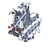
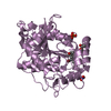
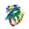
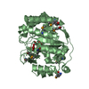
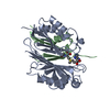
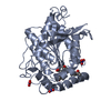
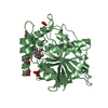
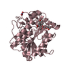
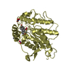
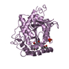
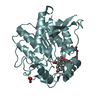
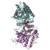
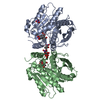
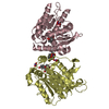
| Title | Crystal Structure of Rice GID1 complexed with GA3 | ||||||
 Components Components | Gibberellin receptor GID1 | ||||||
 Keywords Keywords | HYDROLASE RECEPTOR / alpha/beta hydrolase / lipase / Gibberellin signaling pathway / Hydrolase / Nucleus / Receptor | ||||||
| Function / homology |  Function and homology information Function and homology informationfloral organ morphogenesis / positive regulation of gibberellic acid mediated signaling pathway / gibberellin binding / response to gibberellin / gibberellic acid mediated signaling pathway / Hydrolases / hydrolase activity / nucleus / cytoplasm Similarity search - Function | ||||||
| Biological species |  | ||||||
| Method |  X-RAY DIFFRACTION / SYNCHROTRON / MOLECULAR REPLACEMENT / Resolution: 1.9 Å X-RAY DIFFRACTION / SYNCHROTRON / MOLECULAR REPLACEMENT / Resolution: 1.9 Å | ||||||
 Authors Authors | Shimada, A. / Nakatsu, T. / Ueguchi-Tanaka, M. / Kato, H. / Matsuoka, M. | ||||||
 Citation Citation | Journal: Nature / Year: 2008 Title: Structural basis for gibberellin recognition by its receptor GID1. Authors: Shimada, A. / Ueguchi-Tanaka, M. / Nakatsu, T. / Nakajima, M. / Naoe, Y. / Ohmiya, H. / Kato, H. / Matsuoka, M. | ||||||
| History |
|
- Structure visualization
| Structure viewer | Molecule: MolmilJmol/JSmol |
|---|
- Downloads & links
-Download
| PDBx/mmCIF format | 3ed1.cif.gz | 397.1 KB | Display | PDBx/mmCIF format |
|---|---|---|---|---|
| PDB format | pdb3ed1.ent.gz | 323.7 KB | Display | PDB format |
| PDBx/mmJSON format | 3ed1.json.gz | Tree view | PDBx/mmJSON format | |
| Others |  Other downloads Other downloads |
-Validation report
| Arichive directory | https://data.pdbj.org/pub/pdb/validation_reports/ed/3ed1ftp://data.pdbj.org/pub/pdb/validation_reports/ed/3ed1 | HTTPS FTP |
|---|
-Related structure data
| Related structure data |  3eblSC S: Starting model for refinement C: citing same article ( |
|---|---|
| Similar structure data |

-Links
 PDBj
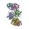
PDBj- Assembly
-Components
-Protein , 1 types, 6 molecules ABCDEF
| #1: Protein | Mass: 40900.699 Da / Num. of mol.: 6 Source method: isolated from a genetically manipulated source Source: (gene. exp.) Gene: GID1, Os05g0407500, LOC_Os05g33730, OJ1657_H11.10, P0040B10.6 Plasmid: pET11b / Production host:  |
|---|
-Non-polymers , 5 types, 1132 molecules 


| #2: Chemical | ChemComp-MPD / (  Mass: 118.174 Da / Num. of mol.: 6 / Source method: obtained synthetically / Formula: C6H14O2 / Comment: precipitant*YM Mass: 118.174 Da / Num. of mol.: 6 / Source method: obtained synthetically / Formula: C6H14O2 / Comment: precipitant*YM#3: Chemical | ChemComp-NO3 /  Mass: 62.005 Da / Num. of mol.: 12 / Source method: obtained synthetically / Formula: NO3 Mass: 62.005 Da / Num. of mol.: 12 / Source method: obtained synthetically / Formula: NO3#4: Chemical | ChemComp-GA3 /  Mass: 346.374 Da / Num. of mol.: 6 / Source method: obtained synthetically / Formula: C19H22O6 Mass: 346.374 Da / Num. of mol.: 6 / Source method: obtained synthetically / Formula: C19H22O6#5: Chemical |  Mass: 94.971 Da / Num. of mol.: 2 / Source method: obtained synthetically / Formula: PO4 Mass: 94.971 Da / Num. of mol.: 2 / Source method: obtained synthetically / Formula: PO4#6: Water | ChemComp-HOH / | Mass: 18.015 Da / Num. of mol.: 1106 / Source method: isolated from a natural source / Formula: H2O |
|---|
-Experimental details
-Experiment
| Experiment | Method: X-RAY DIFFRACTION / Number of used crystals: 1 |
|---|
- Sample preparation
| Crystal | Density Matthews: 2.59 Å3/Da / Density % sol: 52.57 % / Mosaicity: 0.224 ° |
|---|---|
| Crystal grow | Temperature: 293 K / Method: vapor diffusion, sitting drop / pH: 7.5 Details: 15% PEG4000, 8% MPD, 0.2M NaNO3, 0.1M HEPES pH7.5, VAPOR DIFFUSION, SITTING DROP, temperature 293K |
-Data collection
| Diffraction | Mean temperature: 90 K |
|---|---|
| Diffraction source | Source: SYNCHROTRON / Site: SPring-8  / Beamline: BL41XU / Wavelength: 1 Å / Beamline: BL41XU / Wavelength: 1 Å |
| Detector | Type: RAYONIX MX225HE / Detector: CCD / Date: Jul 18, 2008 |
| Radiation | Protocol: SINGLE WAVELENGTH / Monochromatic (M) / Laue (L): M / Scattering type: x-ray |
| Radiation wavelength | Wavelength: 1 Å / Relative weight: 1 |
| Reflection | Resolution: 1.9→50 Å / Num. obs: 192246 / % possible obs: 97.6 % / Observed criterion σ(I): 0 / Redundancy: 3.7 % / Rmerge(I) obs: 0.054 / Χ2: 1.244 / Net I/σ(I): 27.585 |
| Reflection shell | Resolution: 1.9→1.97 Å / Redundancy: 3.3 % / Rmerge(I) obs: 0.365 / Num. unique all: 17227 / Χ2: 1.139 / % possible all: 87.5 |
- Processing
| Software |
| ||||||||||||||||||||||||||||||||||||||||||||||||||||||||||||||||||||||||||||||||||||||||||
|---|---|---|---|---|---|---|---|---|---|---|---|---|---|---|---|---|---|---|---|---|---|---|---|---|---|---|---|---|---|---|---|---|---|---|---|---|---|---|---|---|---|---|---|---|---|---|---|---|---|---|---|---|---|---|---|---|---|---|---|---|---|---|---|---|---|---|---|---|---|---|---|---|---|---|---|---|---|---|---|---|---|---|---|---|---|---|---|---|---|---|---|
| Refinement | Method to determine structure: MOLECULAR REPLACEMENT Starting model: PDB ENTRY 3EBL Resolution: 1.9→20 Å / Cor.coef. Fo:Fc: 0.956 / Cor.coef. Fo:Fc free: 0.935 / WRfactor Rfree: 0.237 / WRfactor Rwork: 0.195 / Occupancy max: 1 / Occupancy min: 0.5 / FOM work R set: 0.822 / SU B: 3.436 / SU ML: 0.101 / SU R Cruickshank DPI: 0.14 / SU Rfree: 0.137 / Cross valid method: THROUGHOUT / σ(F): 0 / ESU R: 0.14 / ESU R Free: 0.137 / Stereochemistry target values: MAXIMUM LIKELIHOOD
| ||||||||||||||||||||||||||||||||||||||||||||||||||||||||||||||||||||||||||||||||||||||||||
| Solvent computation | Ion probe radii: 0.8 Å / Shrinkage radii: 0.8 Å / VDW probe radii: 1.2 Å / Solvent model: BABINET MODEL WITH MASK | ||||||||||||||||||||||||||||||||||||||||||||||||||||||||||||||||||||||||||||||||||||||||||
| Displacement parameters | Biso max: 99.85 Å2 / Biso mean: 31.091 Å2 / Biso min: 12.77 Å2
| ||||||||||||||||||||||||||||||||||||||||||||||||||||||||||||||||||||||||||||||||||||||||||
| Refinement step | Cycle: LAST / Resolution: 1.9→20 Å
| ||||||||||||||||||||||||||||||||||||||||||||||||||||||||||||||||||||||||||||||||||||||||||
| Refine LS restraints |
| ||||||||||||||||||||||||||||||||||||||||||||||||||||||||||||||||||||||||||||||||||||||||||
| LS refinement shell | Resolution: 1.9→1.949 Å / Total num. of bins used: 20
|
