 Movie
Movie Controller

[English] 日本語
 Yorodumi
- PDB-1qz3: CRYSTAL STRUCTURE OF MUTANT M211S/R215L OF CARBOXYLESTERASE EST2 ... -
+ Open data

- Basic information
| Entry | Database: PDB / ID: 1qz3 | ||||||
|---|---|---|---|---|---|---|---|
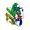
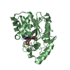
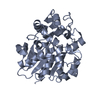
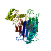
| Title | CRYSTAL STRUCTURE OF MUTANT M211S/R215L OF CARBOXYLESTERASE EST2 COMPLEXED WITH HEXADECANESULFONATE | ||||||
 Components Components | CARBOXYLESTERASE EST2 | ||||||
 Keywords Keywords | HYDROLASE / ALPHA/BETA HYDROLASE FOLD | ||||||
| Function / homology |  Function and homology information Function and homology information | ||||||
| Biological species |  Alicyclobacillus acidocaldarius (bacteria) Alicyclobacillus acidocaldarius (bacteria) | ||||||
| Method |  X-RAY DIFFRACTION / SYNCHROTRON / MOLECULAR REPLACEMENT / Resolution: 2.3 Å X-RAY DIFFRACTION / SYNCHROTRON / MOLECULAR REPLACEMENT / Resolution: 2.3 Å | ||||||
 Authors Authors | De Simone, G. / Mandrich, L. / Menchise, V. / Giordano, V. / Febbraio, F. / Rossi, M. / Pedone, C. / Manco, G. | ||||||
 Citation Citation | Journal: J.Biol.Chem. / Year: 2004 Title: A substrate-induced switch in the reaction mechanism of a thermophilic esterase: kinetic evidences and structural basis. Authors: De Simone, G. / Mandrich, L. / Menchise, V. / Giordano, V. / Febbraio, F. / Rossi, M. / Pedone, C. / Manco, G. #1: Journal: J.Mol.Biol. / Year: 2000Title: A SNAPSHOT OF THE TRANSITION STATE ANALOGUE OF A NOVEL THERMOPHILIC ESTERASE BELONGING TO THE SUBFAMILY OF MAMMALIAN HORMONE-SENSITIVE LIPASE Authors: DE SIMONE, G. / GALDIERO, S. / MANCO, G. / LANG, D. / ROSSI, M. / PEDONE, C. | ||||||
| History |
|
- Structure visualization
| Structure viewer | Molecule: MolmilJmol/JSmol |
|---|
- Downloads & links
-Download
| PDBx/mmCIF format | 1qz3.cif.gz | 78.9 KB | Display | PDBx/mmCIF format |
|---|---|---|---|---|
| PDB format | pdb1qz3.ent.gz | 58.8 KB | Display | PDB format |
| PDBx/mmJSON format | 1qz3.json.gz | Tree view | PDBx/mmJSON format | |
| Others |  Other downloads Other downloads |
-Validation report
| Arichive directory | https://data.pdbj.org/pub/pdb/validation_reports/qz/1qz3ftp://data.pdbj.org/pub/pdb/validation_reports/qz/1qz3 | HTTPS FTP |
|---|
-Related structure data
| Related structure data |  1evqS S: Starting model for refinement |
|---|---|
| Similar structure data |


-Links
 PDBj
PDBj- Assembly
| Deposited unit | 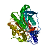
| ||||||||
|---|---|---|---|---|---|---|---|---|---|
| 1 |
| ||||||||
| Unit cell |
|
-Components
| #1: Protein | Mass: 34250.820 Da / Num. of mol.: 1 / Mutation: R215L, M211S Source method: isolated from a genetically manipulated source Source: (gene. exp.) Alicyclobacillus acidocaldarius (bacteria)Plasmid: PT7-7SCII / Species (production host): Escherichia coli / Production host: |
|---|---|
| #2: Chemical | ChemComp-HDS /   Mass: 306.504 Da / Num. of mol.: 1 / Source method: obtained synthetically / Formula: C16H34O3S Mass: 306.504 Da / Num. of mol.: 1 / Source method: obtained synthetically / Formula: C16H34O3S |
| #3: Water | ChemComp-HOH /  Mass: 18.015 Da / Num. of mol.: 253 / Source method: isolated from a natural source / Formula: H2O Mass: 18.015 Da / Num. of mol.: 253 / Source method: isolated from a natural source / Formula: H2O |
| Has protein modification | Y |
-Experimental details
-Experiment
| Experiment | Method: X-RAY DIFFRACTION / Number of used crystals: 1 |
|---|
- Sample preparation
| Crystal | Density Matthews: 2.74 Å3/Da / Density % sol: 55.13 % | ||||||||||||||||||||||||||||||||||||
|---|---|---|---|---|---|---|---|---|---|---|---|---|---|---|---|---|---|---|---|---|---|---|---|---|---|---|---|---|---|---|---|---|---|---|---|---|---|
| Crystal grow | Temperature: 298 K / Method: vapor diffusion, hanging drop / pH: 8.5 Details: AMMONIUM SULPHATE, TRIS BUFFER, pH 8.5, VAPOR DIFFUSION, HANGING DROP, temperature 298K | ||||||||||||||||||||||||||||||||||||
| Crystal grow | *PLUS Temperature: 25 ℃ / Method: vapor diffusion, hanging drop | ||||||||||||||||||||||||||||||||||||
| Components of the solutions | *PLUS
|
-Data collection
| Diffraction | Mean temperature: 100 K |
|---|---|
| Diffraction source | Source: SYNCHROTRON / Site: EMBL/DESY, HAMBURG  / Beamline: X13 / Wavelength: 0.8015 Å / Beamline: X13 / Wavelength: 0.8015 Å |
| Detector | Type: MARRESEARCH / Detector: IMAGE PLATE / Date: Feb 15, 2001 |
| Radiation | Protocol: SINGLE WAVELENGTH / Monochromatic (M) / Laue (L): M / Scattering type: x-ray |
| Radiation wavelength | Wavelength: 0.8015 Å / Relative weight: 1 |
| Reflection | Resolution: 2.3→20 Å / Num. obs: 17707 / % possible obs: 98.8 % / Rsym value: 0.089 / Net I/σ(I): 14.4 |
| Reflection shell | Resolution: 2.3→2.34 Å / Mean I/σ(I) obs: 2.2 / Rsym value: 0.426 / % possible all: 94.1 |
| Reflection | *PLUS Num. measured all: 177515 / Rmerge(I) obs: 0.089 |
| Reflection shell | *PLUS % possible obs: 94.1 % / Rmerge(I) obs: 0.426 |
- Processing
| Software |
| ||||||||||||||||||||||||||||
|---|---|---|---|---|---|---|---|---|---|---|---|---|---|---|---|---|---|---|---|---|---|---|---|---|---|---|---|---|---|
| Refinement | Method to determine structure: MOLECULAR REPLACEMENT Starting model: PDB ENTRY 1EVQ Resolution: 2.3→20 Å / Cross valid method: THROUGHOUT / σ(F): 0 / Stereochemistry target values: Engh & Huber
| ||||||||||||||||||||||||||||
| Refinement step | Cycle: LAST / Resolution: 2.3→20 Å
| ||||||||||||||||||||||||||||
| Refine LS restraints |
| ||||||||||||||||||||||||||||
| Xplor file |
| ||||||||||||||||||||||||||||
| Refinement | *PLUS % reflection Rfree: 5 % | ||||||||||||||||||||||||||||
| Solvent computation | *PLUS | ||||||||||||||||||||||||||||
| Displacement parameters | *PLUS |
