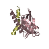
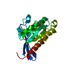
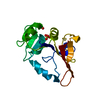
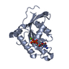
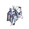
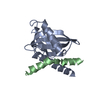
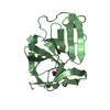
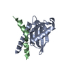
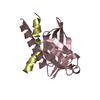
[English] 日本語
Yorodumi
- PDB-3dxc: Crystal structure of the intracellular domain of human APP in com... -

+
Open data


ID or keywords:

Loading...

-
Basic information

Entry
Database: PDB / ID: 3dxc
TitleCrystal structure of the intracellular domain of human APP in complex with Fe65-PTB2
Components
  • Amyloid beta A4 protein
  • Amyloid beta A4 protein-binding family B member 1
KeywordsPROTEIN BINDING / Alzheimer's disease / APP / AICD / Fe65 / PTB domain / Alzheimer disease / Amyloid / Apoptosis / Cell adhesion / Coated pit / Disease mutation / Endocytosis / Glycoprotein / Heparin-binding / Iron / Membrane / Metal-binding / Notch signaling pathway / Phosphoprotein / Protease inhibitor / Proteoglycan / Serine protease inhibitor / Transmembrane
Function / homology
Function and homology information


negative regulation of cell cycle G1/S phase transition / regulation of epidermal growth factor-activated receptor activity / signaling receptor activator activity / collateral sprouting in absence of injury / cytosolic mRNA polyadenylation / microglia development / regulation of synapse structure or activity / Formyl peptide receptors bind formyl peptides and many other ligands / axo-dendritic transport / synaptic assembly at neuromuscular junction ...negative regulation of cell cycle G1/S phase transition / regulation of epidermal growth factor-activated receptor activity / signaling receptor activator activity / collateral sprouting in absence of injury / cytosolic mRNA polyadenylation / microglia development / regulation of synapse structure or activity / Formyl peptide receptors bind formyl peptides and many other ligands / axo-dendritic transport / synaptic assembly at neuromuscular junction / smooth endoplasmic reticulum calcium ion homeostasis / axon midline choice point recognition / astrocyte activation involved in immune response / proline-rich region binding / regulation of spontaneous synaptic transmission / regulation of Wnt signaling pathway / mating behavior / ciliary rootlet / Lysosome Vesicle Biogenesis / PTB domain binding / Golgi-associated vesicle / neuron remodeling / positive regulation of amyloid fibril formation / low-density lipoprotein particle receptor binding / Insertion of tail-anchored proteins into the endoplasmic reticulum membrane / : / Deregulated CDK5 triggers multiple neurodegenerative pathways in Alzheimer's disease models / nuclear envelope lumen / presynaptic active zone / modulation of excitatory postsynaptic potential / suckling behavior / COPII-coated ER to Golgi transport vesicle / dendrite development / smooth endoplasmic reticulum / regulation of NMDA receptor activity / TRAF6 mediated NF-kB activation / Advanced glycosylation endproduct receptor signaling / neuromuscular process controlling balance / The NLRP3 inflammasome / regulation of presynapse assembly / intracellular copper ion homeostasis / transition metal ion binding / regulation of multicellular organism growth / negative regulation of long-term synaptic potentiation / negative regulation of neuron differentiation / smooth muscle contraction / ECM proteoglycans / spindle midzone / positive regulation of T cell migration / Purinergic signaling in leishmaniasis infection / positive regulation of calcium-mediated signaling / forebrain development / regulation of peptidyl-tyrosine phosphorylation / positive regulation of chemokine production / clathrin-coated pit / Notch signaling pathway / positive regulation of G2/M transition of mitotic cell cycle / ionotropic glutamate receptor signaling pathway / positive regulation of protein metabolic process / neuron projection maintenance / cholesterol metabolic process / positive regulation of glycolytic process / extracellular matrix organization / positive regulation of mitotic cell cycle / response to interleukin-1 / axonogenesis / adult locomotory behavior / trans-Golgi network membrane / locomotory behavior / dendritic shaft / platelet alpha granule lumen / positive regulation of peptidyl-threonine phosphorylation / learning / positive regulation of interleukin-1 beta production / central nervous system development / positive regulation of long-term synaptic potentiation / astrocyte activation / endosome lumen / positive regulation of protein secretion / synapse organization / microglial cell activation / Post-translational protein phosphorylation / regulation of long-term neuronal synaptic plasticity / positive regulation of JNK cascade / TAK1-dependent IKK and NF-kappa-B activation / neuromuscular junction / visual learning / serine-type endopeptidase inhibitor activity / recycling endosome / cognition / positive regulation of neuron projection development / positive regulation of inflammatory response / neuron cellular homeostasis / Golgi lumen / endocytosis / positive regulation of interleukin-6 production / positive regulation of non-canonical NF-kappaB signal transduction / cellular response to amyloid-beta / Regulation of Insulin-like Growth Factor (IGF) transport and uptake by Insulin-like Growth Factor Binding Proteins (IGFBPs) / neuron projection development
Similarity search - Function
Amyloid beta precursor protein binding family B member 1/2/3 / Phosphotyrosine interaction domain (PTB/PID) / Phosphotyrosine interaction domain (PID) profile. / Phosphotyrosine-binding domain, phosphotyrosine-interaction (PI) domain / PTB/PI domain / Amyloidogenic glycoprotein, copper-binding / Amyloidogenic glycoprotein, copper-binding domain conserved site / Amyloidogenic glycoprotein, copper-binding domain superfamily / Copper-binding of amyloid precursor, CuBD / Amyloid precursor protein (APP) copper-binding (CuBD) domain signature. ...Amyloid beta precursor protein binding family B member 1/2/3 / Phosphotyrosine interaction domain (PTB/PID) / Phosphotyrosine interaction domain (PID) profile. / Phosphotyrosine-binding domain, phosphotyrosine-interaction (PI) domain / PTB/PI domain / Amyloidogenic glycoprotein, copper-binding / Amyloidogenic glycoprotein, copper-binding domain conserved site / Amyloidogenic glycoprotein, copper-binding domain superfamily / Copper-binding of amyloid precursor, CuBD / Amyloid precursor protein (APP) copper-binding (CuBD) domain signature. / Amyloidogenic glycoprotein, amyloid-beta peptide superfamily / Beta-amyloid peptide (beta-APP) / Amyloidogenic glycoprotein, amyloid-beta peptide / Beta-amyloid precursor protein C-terminal / Amyloidogenic glycoprotein, intracellular domain, conserved site / Beta-amyloid precursor protein C-terminus / Amyloid precursor protein (APP) intracellular domain signature. / Amyloid precursor protein (APP) E1 domain profile. / Amyloid precursor protein (APP) E2 domain profile. / Amyloidogenic glycoprotein, extracellular / Amyloidogenic glycoprotein, heparin-binding / Amyloidogenic glycoprotein, E2 domain / E2 domain superfamily / Amyloidogenic glycoprotein, heparin-binding domain superfamily / Amyloid A4 N-terminal heparin-binding / E2 domain of amyloid precursor protein / amyloid A4 / Amyloidogenic glycoprotein / Proteinase inhibitor I2, Kunitz, conserved site / Pancreatic trypsin inhibitor (Kunitz) family signature. / BPTI/Kunitz family of serine protease inhibitors. / Pancreatic trypsin inhibitor Kunitz domain / Kunitz/Bovine pancreatic trypsin inhibitor domain / Pancreatic trypsin inhibitor (Kunitz) family profile. / Pancreatic trypsin inhibitor Kunitz domain superfamily / WW domain / WW/rsp5/WWP domain signature. / WW domain superfamily / WW/rsp5/WWP domain profile. / Domain with 2 conserved Trp (W) residues / WW domain / Pleckstrin-homology domain (PH domain)/Phosphotyrosine-binding domain (PTB) / PH-domain like / PH-like domain superfamily / Roll / Mainly Beta
Similarity search - Domain/homology
Amyloid beta precursor protein binding family B member 1 / Amyloid-beta precursor protein
Similarity search - Component
Biological speciesHomo sapiens (human)
MethodX-RAY DIFFRACTION / SYNCHROTRON / MOLECULAR REPLACEMENT / Resolution: 2.1 Å
AuthorsRadzimanowski, J. / Sinning, I. / Wild, K.
Citation
Journal: Embo Rep. / Year: 2008
Title: Structure of the intracellular domain of the amyloid precursor protein in complex with Fe65-PTB2.
Authors: Radzimanowski, J. / Simon, B. / Sattler, M. / Beyreuther, K. / Sinning, I. / Wild, K.
#1: Journal: Acta Crystallogr.,Sect.F / Year: 2008
Title: Overproduction, purification, crystallization and preliminary X-ray analysis of human Fe65-PTB2 in complex with the amyloid precursor protein intracellular domain
Authors: Radzimanowski, R. / Beyreuther, K. / Sinning, I. / Wild, K.
History
DepositionJul 24, 2008Deposition site: RCSB / Processing site: RCSB
Revision 1.0Sep 16, 2008Provider: repository / Type: Initial release
Revision 1.1Jul 13, 2011Group: Version format compliance
Revision 1.2Feb 1, 2017Group: Database references
Revision 1.3Feb 21, 2024Group: Data collection / Database references
Category: chem_comp_atom / chem_comp_bond ...chem_comp_atom / chem_comp_bond / database_2 / struct_ref_seq_dif
Item: _database_2.pdbx_DOI / _database_2.pdbx_database_accession / _struct_ref_seq_dif.details

-
Structure visualization

Structure viewerMolecule:
MolmilJmol/JSmol

Downloads & links

-
Assembly

Deposited unit
A: Amyloid beta A4 protein-binding family B member 1
B: Amyloid beta A4 protein
C: Amyloid beta A4 protein-binding family B member 1
D: Amyloid beta A4 protein


Theoretical massNumber of molelcules
Total (without water)39,1104
Polymers39,1104
Non-polymers00
Water4,252236
1
A: Amyloid beta A4 protein-binding family B member 1
B: Amyloid beta A4 protein


Theoretical massNumber of molelcules
Total (without water)19,5552
Polymers19,5552
Non-polymers00
Water362
TypeNameSymmetry operationNumber
identity operation1_555x,y,z1
Buried area2100 Å2
ΔGint-16 kcal/mol
Surface area8190 Å2
MethodPISA
2
C: Amyloid beta A4 protein-binding family B member 1
D: Amyloid beta A4 protein


Theoretical massNumber of molelcules
Total (without water)19,5552
Polymers19,5552
Non-polymers00
Water362
TypeNameSymmetry operationNumber
identity operation1_555x,y,z1
Buried area2120 Å2
ΔGint-13 kcal/mol
Surface area7690 Å2
MethodPISA
Unit cell
Length a, b, c (Å)114.320, 114.320, 74.850
Angle α, β, γ (deg.)90.00, 90.00, 120.00
Int Tables number169
Space group name H-MP61
DetailsTHE ASYMMETRIC UNIT CONTAINS TWO COPIES OF A PROTEIN PROTEIN COMPLEX (ONE BIOMOLECULE 1 BINDS ONE BIOMOLECULE 2).

-
Components

#1: Protein Amyloid beta A4 protein-binding family B member 1


Mass: 15459.690 Da / Num. of mol.: 2 / Fragment: PTB2 domain, UNP residues 534-667
Source method: isolated from a genetically manipulated source
Source: (gene. exp.) Homo sapiens (human) / Gene: APBB1, FE65, RIR / Plasmid: pET21d / Production host: Escherichia coli (E. coli) / Strain (production host): BL21(DE3) RIL / References: UniProt: O00213
#2: Protein/peptide Amyloid beta A4 protein


Mass: 4095.505 Da / Num. of mol.: 2 / Fragment: APP intracellular domain, UNP residues 739-770
Source method: isolated from a genetically manipulated source
Source: (gene. exp.) Homo sapiens (human) / Gene: APP, A4, AD1 / Plasmid: pETtrx_1b / Production host: Escherichia coli (E. coli) / Strain (production host): BL21(DE3) RIL / References: UniProt: P05067
#3: Water ChemComp-HOH / water / Water


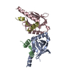
Mass: 18.015 Da / Num. of mol.: 236 / Source method: isolated from a natural source / Formula: H2O

-
Experimental details

-
Experiment

ExperimentMethod: X-RAY DIFFRACTION / Number of used crystals: 1

-
Sample preparation

CrystalDensity Matthews: 3.61 Å3/Da / Density % sol: 65.93 %
Crystal growTemperature: 293 K / Method: vapor diffusion / pH: 4.6
Details: 3.2 M NaCl,0.1 M sodium acetate , pH 4.6, VAPOR DIFFUSION, temperature 293K

-
Data collection

DiffractionMean temperature: 100 K
Diffraction sourceSource: SYNCHROTRON / Site: ESRF / Beamline: ID14-2 / Wavelength: 1 Å
DetectorType: ADSC QUANTUM 4 / Detector: CCD / Date: Jul 19, 2007
RadiationProtocol: SINGLE WAVELENGTH / Monochromatic (M) / Laue (L): M / Scattering type: x-ray
Radiation wavelengthWavelength: 1 Å / Relative weight: 1
ReflectionResolution: 2.1→33 Å / Num. obs: 32612 / % possible obs: 100 % / Redundancy: 11.1 % / Rmerge(I) obs: 0.06 / Rsym value: 0.06 / Net I/σ(I): 28.8
Reflection shellResolution: 2.1→2.21 Å / Redundancy: 11.2 % / Rmerge(I) obs: 0.465 / Mean I/σ(I) obs: 4.7 / Rsym value: 0.465

-
Processing

Software
NameVersionClassification
REFMAC5.3.0008refinement
DNAdata collection
MOSFLMdata reduction
SCALAdata scaling
PHASERphasing
RefinementMethod to determine structure: MOLECULAR REPLACEMENT / Resolution: 2.1→33 Å / Cor.coef. Fo:Fc: 0.951 / Cor.coef. Fo:Fc free: 0.935 / SU B: 4.055 / SU ML: 0.109 / Cross valid method: THROUGHOUT / ESU R: 0.158 / ESU R Free: 0.15 / Stereochemistry target values: MAXIMUM LIKELIHOOD / Details: HYDROGENS HAVE BEEN ADDED IN THE RIDING POSITIONS
RfactorNum. reflection% reflectionSelection details
Rfree0.23816 1997 6.1 %RANDOM
Rwork0.20229 ---
obs0.20449 30579 100 %-
Solvent computationIon probe radii: 0.8 Å / Shrinkage radii: 0.8 Å / VDW probe radii: 1.4 Å / Solvent model: MASK
Displacement parametersBiso mean: 41.32 Å2
Baniso -1Baniso -2Baniso -3
1--0.07 Å2-0.03 Å20 Å2
2---0.07 Å20 Å2
3---0.1 Å2
Refinement stepCycle: LAST / Resolution: 2.1→33 Å
ProteinNucleic acidLigandSolventTotal
Num. atoms2395 0 0 236 2631
Refine LS restraints
Refine-IDTypeDev idealDev ideal targetNumber
X-RAY DIFFRACTIONr_bond_refined_d0.0130.0222477
X-RAY DIFFRACTIONr_bond_other_d
X-RAY DIFFRACTIONr_angle_refined_deg1.3121.9323366
X-RAY DIFFRACTIONr_angle_other_deg
X-RAY DIFFRACTIONr_dihedral_angle_1_deg6.3775307
X-RAY DIFFRACTIONr_dihedral_angle_2_deg34.83223.739115
X-RAY DIFFRACTIONr_dihedral_angle_3_deg14.2115390
X-RAY DIFFRACTIONr_dihedral_angle_4_deg14.3961514
X-RAY DIFFRACTIONr_chiral_restr0.0890.2371
X-RAY DIFFRACTIONr_gen_planes_refined0.0050.021908
X-RAY DIFFRACTIONr_gen_planes_other
X-RAY DIFFRACTIONr_nbd_refined0.2060.21144
X-RAY DIFFRACTIONr_nbd_other
X-RAY DIFFRACTIONr_nbtor_refined0.2970.21697
X-RAY DIFFRACTIONr_nbtor_other
X-RAY DIFFRACTIONr_xyhbond_nbd_refined0.1820.2190
X-RAY DIFFRACTIONr_xyhbond_nbd_other
X-RAY DIFFRACTIONr_metal_ion_refined
X-RAY DIFFRACTIONr_metal_ion_other
X-RAY DIFFRACTIONr_symmetry_vdw_refined0.2190.247
X-RAY DIFFRACTIONr_symmetry_vdw_other
X-RAY DIFFRACTIONr_symmetry_hbond_refined0.1910.218
X-RAY DIFFRACTIONr_symmetry_hbond_other
X-RAY DIFFRACTIONr_symmetry_metal_ion_refined
X-RAY DIFFRACTIONr_symmetry_metal_ion_other
X-RAY DIFFRACTIONr_mcbond_it1.0031.51585
X-RAY DIFFRACTIONr_mcbond_other
X-RAY DIFFRACTIONr_mcangle_it1.7122490
X-RAY DIFFRACTIONr_scbond_it2.18831008
X-RAY DIFFRACTIONr_scangle_it3.234.5876
X-RAY DIFFRACTIONr_rigid_bond_restr
X-RAY DIFFRACTIONr_sphericity_free
X-RAY DIFFRACTIONr_sphericity_bonded
LS refinement shellResolution: 2.1→2.154 Å / Total num. of bins used: 20
RfactorNum. reflection% reflection
Rfree0.312 142 -
Rwork0.27 2234 -
obs--100 %

+
About Yorodumi

-
News

-
Feb 9, 2022. New format data for meta-information of EMDB entries

New format data for meta-information of EMDB entries

  • Version 3 of the EMDB header file is now the official format.
  • The previous official version 1.9 will be removed from the archive.

Related info.:EMDB header

External links:wwPDB to switch to version 3 of the EMDB data model

-
Aug 12, 2020. Covid-19 info

Covid-19 info

URL: https://pdbj.org/emnavi/covid19.php

New page: Covid-19 featured information page in EM Navigator.

Related info.:Covid-19 info / Mar 5, 2020. Novel coronavirus structure data

+
Mar 5, 2020. Novel coronavirus structure data

Novel coronavirus structure data

Related info.:Yorodumi Speices / Aug 12, 2020. Covid-19 info

External links:COVID-19 featured content - PDBj / Molecule of the Month (242):Coronavirus Proteases

+
Jan 31, 2019. EMDB accession codes are about to change! (news from PDBe EMDB page)

EMDB accession codes are about to change! (news from PDBe EMDB page)

  • The allocation of 4 digits for EMDB accession codes will soon come to an end. Whilst these codes will remain in use, new EMDB accession codes will include an additional digit and will expand incrementally as the available range of codes is exhausted. The current 4-digit format prefixed with “EMD-” (i.e. EMD-XXXX) will advance to a 5-digit format (i.e. EMD-XXXXX), and so on. It is currently estimated that the 4-digit codes will be depleted around Spring 2019, at which point the 5-digit format will come into force.
  • The EM Navigator/Yorodumi systems omit the EMD- prefix.

Related info.:Q: What is EMD? / ID/Accession-code notation in Yorodumi/EM Navigator

External links:EMDB Accession Codes are Changing Soon! / Contact to PDBj

+
Jul 12, 2017. Major update of PDB

Major update of PDB

  • wwPDB released updated PDB data conforming to the new PDBx/mmCIF dictionary.
  • This is a major update changing the version number from 4 to 5, and with Remediation, in which all the entries are updated.
  • In this update, many items about electron microscopy experimental information are reorganized (e.g. em_software).
  • Now, EM Navigator and Yorodumi are based on the updated data.

External links:wwPDB Remediation / Enriched Model Files Conforming to OneDep Data Standards Now Available in the PDB FTP Archive

-
Yorodumi

Thousand views of thousand structures

  • Yorodumi is a browser for structure data from EMDB, PDB, SASBDB, etc.
  • This page is also the successor to EM Navigator detail page, and also detail information page/front-end page for Omokage search.
  • The word "yorodu" (or yorozu) is an old Japanese word meaning "ten thousand". "mi" (miru) is to see.

Related info.:EMDB / PDB / SASBDB / Comparison of 3 databanks / Yorodumi Search / Aug 31, 2016. New EM Navigator & Yorodumi / Yorodumi Papers / Jmol/JSmol / Function and homology information / Changes in new EM Navigator and Yorodumi

Read more