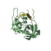
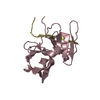
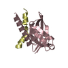
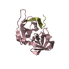
[English] 日本語
Yorodumi
- PDB-2qv1: Crystal structure of HCV NS3-4A V36M mutant -

+
Open data


ID or keywords:

Loading...

-
Basic information

Entry
Database: PDB / ID: 2qv1
TitleCrystal structure of HCV NS3-4A V36M mutant
Components
  • NS3
  • peptide
KeywordsHYDROLASE / HCV / protease / NS3-4A / V36M mutant
Function / homology
Function and homology information


positive regulation of hexokinase activity / modulation by virus of host cellular process / translocation of peptides or proteins into host cell cytoplasm / Toll-like receptor 2 binding / viral capsid assembly / adhesion receptor-mediated virion attachment to host cell / TBC/RABGAPs / hepacivirin / host cell mitochondrial membrane / host cell lipid droplet ...positive regulation of hexokinase activity / modulation by virus of host cellular process / translocation of peptides or proteins into host cell cytoplasm / Toll-like receptor 2 binding / viral capsid assembly / adhesion receptor-mediated virion attachment to host cell / TBC/RABGAPs / hepacivirin / host cell mitochondrial membrane / host cell lipid droplet / symbiont-mediated suppression of host TRAF-mediated signal transduction / transformation of host cell by virus / positive regulation of cytokinesis / symbiont-mediated perturbation of host cell cycle G1/S transition checkpoint / negative regulation of protein secretion / symbiont-mediated suppression of host JAK-STAT cascade via inhibition of STAT1 activity / symbiont-mediated suppression of host cytoplasmic pattern recognition receptor signaling pathway via inhibition of MAVS activity / endoplasmic reticulum-Golgi intermediate compartment membrane / serine-type peptidase activity / ribonucleoside triphosphate phosphatase activity / kinase binding / SH3 domain binding / : / viral capsid / nucleoside-triphosphate phosphatase / protein complex oligomerization / monoatomic ion channel activity / clathrin-dependent endocytosis of virus by host cell / viral nucleocapsid / Hydrolases; Acting on peptide bonds (peptidases); Cysteine endopeptidases / RNA helicase activity / entry receptor-mediated virion attachment to host cell / host cell endoplasmic reticulum membrane / membrane => GO:0016020 / host cell perinuclear region of cytoplasm / RNA helicase / induction by virus of host autophagy / RNA-directed RNA polymerase / symbiont entry into host cell / ribonucleoprotein complex / viral RNA genome replication / cysteine-type endopeptidase activity / RNA-dependent RNA polymerase activity / serine-type endopeptidase activity / fusion of virus membrane with host endosome membrane / viral envelope / symbiont-mediated suppression of host type I interferon-mediated signaling pathway / host cell nucleus / structural molecule activity / virion attachment to host cell / endoplasmic reticulum membrane / host cell plasma membrane / virion membrane / negative regulation of transcription by RNA polymerase II / ATP hydrolysis activity / proteolysis / RNA binding / zinc ion binding / ATP binding / membrane / metal ion binding
Similarity search - Function
Thrombin, subunit H - #120 / Hepatitis C virus core protein, chain A superfamily / Hepatitus C virus, Non-structural 5a protein, C-terminal / Hepatitis C virus NS5A, 1B domain superfamily / Hepatitis C virus non-structural protein NS2, C-terminal domain / Hepatitis C virus non-structural protein NS2, N-terminal domain / Hepatitis C virus non-structural protein NS2 / HCV NS5a protein C-terminal region / Hepatitis C virus, Non-structural protein NS4b / Hepatitis C virus, Core protein, N-terminal ...Thrombin, subunit H - #120 / Hepatitis C virus core protein, chain A superfamily / Hepatitus C virus, Non-structural 5a protein, C-terminal / Hepatitis C virus NS5A, 1B domain superfamily / Hepatitis C virus non-structural protein NS2, C-terminal domain / Hepatitis C virus non-structural protein NS2, N-terminal domain / Hepatitis C virus non-structural protein NS2 / HCV NS5a protein C-terminal region / Hepatitis C virus, Non-structural protein NS4b / Hepatitis C virus, Core protein, N-terminal / Hepatitis C virus non-structural protein NS4b / Hepatitis C virus capsid protein / Hepatitis C virus, Non-structural protein NS2 / Hepatitis C virus, Non-structural 5a protein / Hepatitis C virus, Non-structural 5a protein, domain 1a / Hepatitis C virus non-structural 5a, 1B domain / NS5A domain 1a superfamily / Hepatitis C virus non-structural 5a protein membrane anchor / Hepatitis C virus non-structural 5a zinc finger domain / Hepatitis C virus non-structural 5a domain 1b / Hepacivirus nonstructural protein 2 (NS2) protease domain profile. / Hepatitis C virus, Non-structural protein NS4a / Hepatitis C virus non-structural protein NS4a / Hepatitis C virus, Core protein, C-terminal / Hepatitis C virus core protein / Hepatitis C virus, Non-structural protein E2/NS1 / Hepatitis C virus non-structural protein E2/NS1 / Hepatitis C virus, Envelope glycoprotein E1 / Hepatitis C virus envelope glycoprotein E1 / RNA dependent RNA polymerase, hepatitis C virus / Viral RNA dependent RNA polymerase / Hepatitis C virus, NS3 protease, Peptidase S29 / Hepatitis C virus NS3 protease / Hepacivirus/Pegivirus NS3 protease domain profile. / DEAD box, Flavivirus / Flavivirus DEAD domain / helicase superfamily c-terminal domain / Superfamilies 1 and 2 helicase C-terminal domain profile. / Superfamilies 1 and 2 helicase ATP-binding type-1 domain profile. / DEAD-like helicases superfamily / Helicase, C-terminal / Helicase superfamily 1/2, ATP-binding domain / Trypsin-like serine proteases / Thrombin, subunit H / Reverse transcriptase/Diguanylate cyclase domain / RNA-directed RNA polymerase, catalytic domain / RdRp of positive ssRNA viruses catalytic domain profile. / Peptidase S1, PA clan, chymotrypsin-like fold / Peptidase S1, PA clan / DNA/RNA polymerase superfamily / Beta Barrel / P-loop containing nucleoside triphosphate hydrolase / Mainly Beta
Similarity search - Domain/homology
Genome polyprotein / Genome polyprotein / Genome polyprotein
Similarity search - Component
Biological speciesHepatitis C virus
MethodX-RAY DIFFRACTION / SYNCHROTRON / FOURIER SYNTHESIS / Resolution: 2.4 Å
AuthorsWei, Y.
CitationJournal: To be Published
Title: Phenotypic and Structural Analyses of HCV NS3 Protease Val36 Variants
Authors: Wei, Y. / Chao, L.
History
DepositionAug 7, 2007Deposition site: RCSB / Processing site: RCSB
Revision 1.0Aug 19, 2008Provider: repository / Type: Initial release
Revision 1.1Jul 13, 2011Group: Version format compliance
Revision 1.2Sep 9, 2020Group: Database references / Derived calculations / Structure summary
Category: struct_conn / struct_keywords ...struct_conn / struct_keywords / struct_ref_seq_dif / struct_site
Item: _struct_conn.ptnr1_auth_comp_id / _struct_conn.ptnr1_auth_seq_id ..._struct_conn.ptnr1_auth_comp_id / _struct_conn.ptnr1_auth_seq_id / _struct_conn.ptnr1_label_asym_id / _struct_conn.ptnr1_label_atom_id / _struct_conn.ptnr1_label_comp_id / _struct_conn.ptnr1_label_seq_id / _struct_conn.ptnr2_auth_comp_id / _struct_conn.ptnr2_auth_seq_id / _struct_conn.ptnr2_label_asym_id / _struct_conn.ptnr2_label_atom_id / _struct_conn.ptnr2_label_comp_id / _struct_conn.ptnr2_label_seq_id / _struct_keywords.text / _struct_ref_seq_dif.details / _struct_site.pdbx_auth_asym_id / _struct_site.pdbx_auth_comp_id / _struct_site.pdbx_auth_seq_id
Revision 1.3Oct 20, 2021Group: Database references / Category: database_2 / struct_ref_seq_dif
Item: _database_2.pdbx_DOI / _database_2.pdbx_database_accession / _struct_ref_seq_dif.details
Revision 1.4Feb 21, 2024Group: Data collection / Category: chem_comp_atom / chem_comp_bond
Revision 1.5Apr 3, 2024Group: Refinement description / Category: pdbx_initial_refinement_model

-
Structure visualization

Structure viewerMolecule:
MolmilJmol/JSmol

Downloads & links

-
Assembly

Deposited unit
A: NS3
B: NS3
C: peptide
D: peptide
hetero molecules


Theoretical massNumber of molelcules
Total (without water)42,7106
Polymers42,5794
Non-polymers1312
Water4,017223
1
A: NS3
C: peptide
hetero molecules


Theoretical massNumber of molelcules
Total (without water)21,3553
Polymers21,2902
Non-polymers651
Water362
TypeNameSymmetry operationNumber
identity operation1_555x,y,z1
Buried area1700 Å2
ΔGint-13 kcal/mol
Surface area8010 Å2
MethodPISA
2
B: NS3
D: peptide
hetero molecules


Theoretical massNumber of molelcules
Total (without water)21,3553
Polymers21,2902
Non-polymers651
Water362
TypeNameSymmetry operationNumber
identity operation1_555x,y,z1
Buried area2490 Å2
ΔGint-14 kcal/mol
Surface area9880 Å2
MethodPISA
Unit cell
Length a, b, c (Å)228.760, 228.760, 75.530
Angle α, β, γ (deg.)90.00, 90.00, 120.00
Int Tables number155
Space group name H-MH32

-
Components

#1: Protein NS3


Mass: 19212.012 Da / Num. of mol.: 2 / Fragment: HCV protease domain NS3 / Mutation: V36M
Source method: isolated from a genetically manipulated source
Source: (gene. exp.) Hepatitis C virus / Strain: H / Gene: NS3 / Plasmid: pBEV10 / Production host: Escherichia coli (E. coli) / Strain (production host): JM109 / References: UniProt: A1Z094, UniProt: A1Z093*PLUS
#2: Protein/peptide peptide /


Mass: 2077.577 Da / Num. of mol.: 2 / Source method: obtained synthetically / References: UniProt: P27958*PLUS
#3: Chemical ChemComp-ZN / ZINC ION


Mass: 65.409 Da / Num. of mol.: 2 / Source method: obtained synthetically / Formula: Zn
#4: Water ChemComp-HOH / water / Water


Mass: 18.015 Da / Num. of mol.: 223 / Source method: isolated from a natural source / Formula: H2O

-
Experimental details

-
Experiment

ExperimentMethod: X-RAY DIFFRACTION / Number of used crystals: 1

-
Sample preparation

CrystalDensity Matthews: 4.47 Å3/Da / Density % sol: 72.46 %
Crystal growTemperature: 298 K / Method: vapor diffusion, hanging drop / pH: 6.2
Details: 1.4M NaCl, 0.3M KH2PO4, 0.1M MES,10.0mM PME, protein concentration 8mg/ml, pH 6.2, VAPOR DIFFUSION, HANGING DROP, temperature 298K

-
Data collection

DiffractionMean temperature: 100 K
Diffraction sourceSource: SYNCHROTRON / Site: ALS / Beamline: 5.0.1 / Wavelength: 1 Å
DetectorType: ADSC QUANTUM 210 / Detector: CCD / Date: Jul 2, 2006
RadiationProtocol: SINGLE WAVELENGTH / Monochromatic (M) / Laue (L): M / Scattering type: x-ray
Radiation wavelengthWavelength: 1 Å / Relative weight: 1
ReflectionResolution: 2.4→25 Å / Num. all: 28682 / Num. obs: 26385 / % possible obs: 91.2 % / Observed criterion σ(F): 3 / Observed criterion σ(I): 2 / Redundancy: 4.6 % / Rmerge(I) obs: 0.052 / Rsym value: 0.039 / Net I/σ(I): 16.5
Reflection shellResolution: 2.4→2.55 Å / Redundancy: 4.6 % / Rmerge(I) obs: 0.052 / Mean I/σ(I) obs: 16.5 / Num. unique all: 28682 / Rsym value: 0.039 / % possible all: 91.2

-
Processing

Software
NameVersionClassification
CNX2005refinement
ADSCQuantumdata collection
DENZOdata reduction
SCALEPACKdata scaling
CNX2005phasing
RefinementMethod to determine structure: FOURIER SYNTHESIS
Starting model: HCV NS3-4A wild type protease

Resolution: 2.4→19.76 Å / Rfactor Rfree error: 0.007 / Data cutoff high absF: 4080894.96 / Data cutoff low absF: 0 / Isotropic thermal model: RESTRAINED / Cross valid method: THROUGHOUT / σ(F): 2 / Stereochemistry target values: Engh & Huber
RfactorNum. reflection% reflectionSelection details
Rfree0.265 1437 5.1 %RANDOM
Rwork0.228 ---
obs-26385 96.4 %-
Solvent computationSolvent model: FLAT MODEL / Bsol: 55.8641 Å2 / ksol: 0.372254 e/Å3
Displacement parametersBiso mean: 53.7 Å2
Baniso -1Baniso -2Baniso -3
1--5.43 Å2-0.24 Å20 Å2
2---5.43 Å20 Å2
3---10.87 Å2
Refine analyze
FreeObs
Luzzati coordinate error0.39 Å0.34 Å
Luzzati d res low-5 Å
Luzzati sigma a0.5 Å0.45 Å
Refinement stepCycle: LAST / Resolution: 2.4→19.76 Å
ProteinNucleic acidLigandSolventTotal
Num. atoms2690 0 2 223 2915
Refine LS restraints
Refine-IDTypeDev idealDev ideal target
X-RAY DIFFRACTIONc_bond_d0.007
X-RAY DIFFRACTIONc_angle_deg1
X-RAY DIFFRACTIONc_dihedral_angle_d20.1
X-RAY DIFFRACTIONc_improper_angle_d0.73
X-RAY DIFFRACTIONc_mcbond_it1.31.5
X-RAY DIFFRACTIONc_mcangle_it2.312
X-RAY DIFFRACTIONc_scbond_it1.662
X-RAY DIFFRACTIONc_scangle_it2.612.5
LS refinement shellResolution: 2.4→2.55 Å / Rfactor Rfree error: 0.028 / Total num. of bins used: 6
RfactorNum. reflection% reflection
Rfree0.396 207 4.7 %
Rwork0.34 4151 -
obs--89.9 %
Xplor file
Refine-IDSerial noParam fileTopol file
X-RAY DIFFRACTION1protein_rep.pprotein.top
X-RAY DIFFRACTION2water_rep.parwater_rep.top
X-RAY DIFFRACTION3ion.paramion.top

+
About Yorodumi

-
News

-
Feb 9, 2022. New format data for meta-information of EMDB entries

New format data for meta-information of EMDB entries

  • Version 3 of the EMDB header file is now the official format.
  • The previous official version 1.9 will be removed from the archive.

Related info.:EMDB header

External links:wwPDB to switch to version 3 of the EMDB data model

-
Aug 12, 2020. Covid-19 info

Covid-19 info

URL: https://pdbj.org/emnavi/covid19.php

New page: Covid-19 featured information page in EM Navigator.

Related info.:Covid-19 info / Mar 5, 2020. Novel coronavirus structure data

+
Mar 5, 2020. Novel coronavirus structure data

Novel coronavirus structure data

Related info.:Yorodumi Speices / Aug 12, 2020. Covid-19 info

External links:COVID-19 featured content - PDBj / Molecule of the Month (242):Coronavirus Proteases

+
Jan 31, 2019. EMDB accession codes are about to change! (news from PDBe EMDB page)

EMDB accession codes are about to change! (news from PDBe EMDB page)

  • The allocation of 4 digits for EMDB accession codes will soon come to an end. Whilst these codes will remain in use, new EMDB accession codes will include an additional digit and will expand incrementally as the available range of codes is exhausted. The current 4-digit format prefixed with “EMD-” (i.e. EMD-XXXX) will advance to a 5-digit format (i.e. EMD-XXXXX), and so on. It is currently estimated that the 4-digit codes will be depleted around Spring 2019, at which point the 5-digit format will come into force.
  • The EM Navigator/Yorodumi systems omit the EMD- prefix.

Related info.:Q: What is EMD? / ID/Accession-code notation in Yorodumi/EM Navigator

External links:EMDB Accession Codes are Changing Soon! / Contact to PDBj

+
Jul 12, 2017. Major update of PDB

Major update of PDB

  • wwPDB released updated PDB data conforming to the new PDBx/mmCIF dictionary.
  • This is a major update changing the version number from 4 to 5, and with Remediation, in which all the entries are updated.
  • In this update, many items about electron microscopy experimental information are reorganized (e.g. em_software).
  • Now, EM Navigator and Yorodumi are based on the updated data.

External links:wwPDB Remediation / Enriched Model Files Conforming to OneDep Data Standards Now Available in the PDB FTP Archive

-
Yorodumi

Thousand views of thousand structures

  • Yorodumi is a browser for structure data from EMDB, PDB, SASBDB, etc.
  • This page is also the successor to EM Navigator detail page, and also detail information page/front-end page for Omokage search.
  • The word "yorodu" (or yorozu) is an old Japanese word meaning "ten thousand". "mi" (miru) is to see.

Related info.:EMDB / PDB / SASBDB / Comparison of 3 databanks / Yorodumi Search / Aug 31, 2016. New EM Navigator & Yorodumi / Yorodumi Papers / Jmol/JSmol / Function and homology information / Changes in new EM Navigator and Yorodumi

Read more