 Movie
Movie Controller

[English] 日本語
 Yorodumi
- PDB-3eyd: Structure of HCV NS3-4A Protease with an Inhibitor Derived from a... -
+ Open data

- Basic information
| Entry | Database: PDB / ID: 3eyd | ||||||
|---|---|---|---|---|---|---|---|
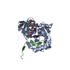
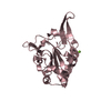
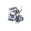
| Title | Structure of HCV NS3-4A Protease with an Inhibitor Derived from a Boronic Acid | ||||||
 Components Components |
| ||||||
 Keywords Keywords | VIRAL PROTEIN / Hepatitis C Virus / NS3 Protease Domain / serine protease / boronic acid inhibitor / Envelope protein / Helicase / Hydrolase / Nucleotide-binding / RNA replication / Transmembrane | ||||||
| Function / homology |  Function and homology information Function and homology informationpositive regulation of metabolic process / regulation of primary metabolic process / hepacivirin / host cell mitochondrial membrane / host cell lipid droplet / symbiont-mediated transformation of host cell / symbiont-mediated suppression of host TRAF-mediated signal transduction / symbiont-mediated perturbation of host cell cycle G1/S transition checkpoint / symbiont-mediated suppression of host JAK-STAT cascade via inhibition of STAT1 activity / symbiont-mediated suppression of host cytoplasmic pattern recognition receptor signaling pathway via inhibition of MAVS activity ...positive regulation of metabolic process / regulation of primary metabolic process / hepacivirin / host cell mitochondrial membrane / host cell lipid droplet / symbiont-mediated transformation of host cell / symbiont-mediated suppression of host TRAF-mediated signal transduction / symbiont-mediated perturbation of host cell cycle G1/S transition checkpoint / symbiont-mediated suppression of host JAK-STAT cascade via inhibition of STAT1 activity / symbiont-mediated suppression of host cytoplasmic pattern recognition receptor signaling pathway via inhibition of MAVS activity / SH3 domain binding / ribonucleoside triphosphate phosphatase activity / nucleoside-triphosphate phosphatase / viral nucleocapsid / channel activity / monoatomic ion transmembrane transport / clathrin-dependent endocytosis of virus by host cell / Hydrolases; Acting on peptide bonds (peptidases); Cysteine endopeptidases / RNA helicase activity / host cell perinuclear region of cytoplasm / host cell endoplasmic reticulum membrane / RNA helicase / symbiont-mediated suppression of host type I interferon-mediated signaling pathway / ribonucleoprotein complex / serine-type endopeptidase activity / viral translational frameshifting / symbiont-mediated activation of host autophagy / RNA-directed RNA polymerase / cysteine-type endopeptidase activity / viral RNA genome replication / RNA-directed RNA polymerase activity / fusion of virus membrane with host endosome membrane / viral envelope / virion attachment to host cell / host cell nucleus / host cell plasma membrane / virion membrane / structural molecule activity / ATP hydrolysis activity / proteolysis / RNA binding / zinc ion binding / ATP binding Similarity search - Function | ||||||
| Biological species |  Hepatitis C virus subtype 1a Hepatitis C virus subtype 1a | ||||||
| Method |  X-RAY DIFFRACTION / SYNCHROTRON / FOURIER SYNTHESIS / Resolution: 2.3 Å X-RAY DIFFRACTION / SYNCHROTRON / FOURIER SYNTHESIS / Resolution: 2.3 Å | ||||||
 Authors Authors | Venkatraman, S. / Wu, W. / Prongay, A.J. / Girijavallabhan, V. / Njoroge, F.G. | ||||||
 Citation Citation | Journal: Bioorg.Med.Chem.Lett. / Year: 2009 Title: Potent inhibitors of HCV-NS3 protease derived from boronic acids. Authors: Venkatraman, S. / Wu, W. / Prongay, A. / Girijavallabhan, V. / George Njoroge, F. | ||||||
| History |
|
- Structure visualization
| Structure viewer | Molecule: MolmilJmol/JSmol |
|---|
- Downloads & links
-Download
| PDBx/mmCIF format | 3eyd.cif.gz | 91.5 KB | Display | PDBx/mmCIF format |
|---|---|---|---|---|
| PDB format | pdb3eyd.ent.gz | 68.2 KB | Display | PDB format |
| PDBx/mmJSON format | 3eyd.json.gz | Tree view | PDBx/mmJSON format | |
| Others |  Other downloads Other downloads |
-Validation report
| Arichive directory | https://data.pdbj.org/pub/pdb/validation_reports/ey/3eydftp://data.pdbj.org/pub/pdb/validation_reports/ey/3eyd | HTTPS FTP |
|---|
-Related structure data
| Similar structure data |
|---|


-Links
 PDBj
PDBj
- Assembly
| Deposited unit | 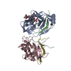
| ||||||||
|---|---|---|---|---|---|---|---|---|---|
| 1 | 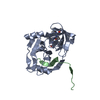
| ||||||||
| 2 | 
| ||||||||
| 3 |
| ||||||||
| Unit cell |
| ||||||||
| Details | The asymmetric unit contains a dimer of the NS3-NS4a complex. This is only the protease domain of NS3 and a peptide of NS4a. This dimeric structure is the result of the soultion structure of the domain. The full length NS3 contains a 3-domain helicase as well and would not have the same dimeric interfaces. |
-Components
| #1: Protein | Mass: 21233.225 Da / Num. of mol.: 2 / Fragment: Protease domain, UNP residues 1027-1207 Source method: isolated from a genetically manipulated source Details: T7 epitope (MASMTGGQQMG) followed by HCV NS3 residues 1-181 the SGHHHHHH Source: (gene. exp.) Hepatitis C virus subtype 1a / Strain: H77 Strain of genotype 1a / Gene: ns3 / Plasmid: pET-3a (Novagen) / Production host:  #2: Protein/peptide | Mass: 2394.039 Da / Num. of mol.: 2 / Fragment: UNP residues 1678-1696 / Mutation: C22S / Source method: obtained synthetically Details: Peptides were synthesized using Fmoc solid-phase chemistry on an ABI 431 synthesizer (Foster City, CA). Preloaded 2-chlorotrityl chloride resin or Wang resin was used for the solid phase ...Details: Peptides were synthesized using Fmoc solid-phase chemistry on an ABI 431 synthesizer (Foster City, CA). Preloaded 2-chlorotrityl chloride resin or Wang resin was used for the solid phase assembly of NS4A activator peptide. The sequence of the peptide was Lys-Lys-Gly-Ser-Val-Val-Ile-Val-Gly-Arg-Ile-Ile-Leu-Ser-Gly-Arg-Pro-Ala-Ile-Val-Pro-Lys-Lys-OH. Trifunctional residue sidechain protecting groups included tert-butyl for Ser, tert-butoxycarbonyl for Lys, and 2,2,4,6,7-pentamethyldihydrobenzofuran-5-sulfonyl for Arg. Cleavage and sidechain deprotection was accomplished using 92.5% trifluoroacetic acid, with 2.5% each of water, ethanedithiol and triisopropylsilane for 2 hours. The peptide was purified by reversed phase HPLC. The peptide molecular weight was confirmed by electrospray ionization mass spectrometry. References: UniProt: Q9ELS8, UniProt: P26664*PLUS #3: Chemical |   Mass: 65.409 Da / Num. of mol.: 2 / Source method: obtained synthetically / Formula: Zn Mass: 65.409 Da / Num. of mol.: 2 / Source method: obtained synthetically / Formula: Zn#4: Chemical | ChemComp-BE8 / [( |   Mass: 627.644 Da / Num. of mol.: 1 / Source method: obtained synthetically / Formula: C29H54BN5O7S Mass: 627.644 Da / Num. of mol.: 1 / Source method: obtained synthetically / Formula: C29H54BN5O7S#5: Water | ChemComp-HOH / |  Mass: 18.015 Da / Num. of mol.: 257 / Source method: isolated from a natural source / Formula: H2O Mass: 18.015 Da / Num. of mol.: 257 / Source method: isolated from a natural source / Formula: H2OHas protein modification | Y | |
|---|
-Experimental details
-Experiment
| Experiment | Method: X-RAY DIFFRACTION / Number of used crystals: 1 |
|---|
- Sample preparation
| Crystal | Density Matthews: 3.89 Å3/Da / Density % sol: 68.37 % |
|---|---|
| Crystal grow | Method: vapor diffusion, hanging drop / pH: 5.6 Details: Crystallization was performed by the vapor diffusion method using hanging drops (4 L protein solution mixed with 4 L (0.75 - 1.00) M NaCl - 0.1 M MES - 0.1 M Na/K PO4, pH 5.6 - 6.2) ...Details: Crystallization was performed by the vapor diffusion method using hanging drops (4 L protein solution mixed with 4 L (0.75 - 1.00) M NaCl - 0.1 M MES - 0.1 M Na/K PO4, pH 5.6 - 6.2) suspended over 1 mL reservoir solutions of (1.25 - 1.50) M NaCl - 0.1 M MES - 0.1 M Na/K PO4 - 5 mM -mercaptoethanol, pH 5.6-6.2. The trays were set at 4oC for 5-7 days to control nucleation, followed by incubation for 3 weeks at 12oC to maximize crystal growth., VAPOR DIFFUSION, HANGING DROP, temperature 277, then 285K Temp details: 277, then 285 |
-Data collection
| Diffraction | Mean temperature: 100 K |
|---|---|
| Diffraction source | Source: SYNCHROTRON / Site: APS  / Beamline: 17-ID / Wavelength: 1 Å / Beamline: 17-ID / Wavelength: 1 Å |
| Detector | Type: ADSC QUANTUM 210 / Detector: CCD / Date: Apr 7, 2004 |
| Radiation | Protocol: SINGLE WAVELENGTH / Monochromatic (M) / Laue (L): M / Scattering type: x-ray |
| Radiation wavelength | Wavelength: 1 Å / Relative weight: 1 |
| Reflection | Resolution: 2.15→50 Å / Num. all: 39911 / Num. obs: 28576 / % possible obs: 71.6 % / Observed criterion σ(I): 1 / Redundancy: 2.8 % / Rmerge(I) obs: 0.075 / Net I/σ(I): 13.5 |
| Reflection shell | Resolution: 2.15→2.2 Å / Rmerge(I) obs: 0.288 / Mean I/σ(I) obs: 2 / Num. unique all: 2631 / % possible all: 20.6 |
- Processing
| Software |
| |||||||||||||||||||||
|---|---|---|---|---|---|---|---|---|---|---|---|---|---|---|---|---|---|---|---|---|---|---|
| Refinement | Method to determine structure: FOURIER SYNTHESIS Starting model: HCV NS3(1-181)S139A - NS4a pdb2o8m.ent Resolution: 2.3→8 Å / Isotropic thermal model: anoistropic / Cross valid method: THROUGHOUT / σ(F): 1
| |||||||||||||||||||||
| Refinement step | Cycle: LAST / Resolution: 2.3→8 Å
| |||||||||||||||||||||
| Refine LS restraints |
| |||||||||||||||||||||
| LS refinement shell | Resolution: 2.3→2.4 Å
|
