 Movie
Movie Controller

[English] 日本語
 Yorodumi
- PDB-3du1: The 2.0 Angstrom Resolution Crystal Structure of HetL, a Pentapep... -
+ Open data

- Basic information
| Entry | Database: PDB / ID: 3du1 | ||||||
|---|---|---|---|---|---|---|---|
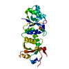
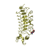
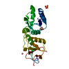
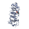
| Title | The 2.0 Angstrom Resolution Crystal Structure of HetL, a Pentapeptide Repeat Protein involved in Heterocyst Differentiation Regulation from the Cyanobacterium Nostoc sp. Strain PCC 7120 | ||||||
 Components Components | All3740 protein | ||||||
 Keywords Keywords | STRUCTURAL PROTEIN / Right-handed Beta Helix / Repeated Five Residue Fold / Pentapeptide Repeat Protein | ||||||
| Function / homology | Pentapeptide repeats (8 copies) / Pentapeptide repeat / E3 ubiquitin-protein ligase SopA / Pectate Lyase C-like / 3 Solenoid / Mainly Beta / All3740 protein Function and homology information Function and homology information | ||||||
| Biological species |  Nostoc sp. (bacteria) Nostoc sp. (bacteria) | ||||||
| Method |  X-RAY DIFFRACTION / sulfur anomalous scattering / Resolution: 2 Å X-RAY DIFFRACTION / sulfur anomalous scattering / Resolution: 2 Å | ||||||
 Authors Authors | Kennedy, M.A. / Ni, S. / Sheldrick, G.M. | ||||||
 Citation Citation | Journal: J.Struct.Biol. / Year: 2008 Title: The 2A resolution crystal structure of HetL, a pentapeptide repeat protein involved in regulation of heterocyst differentiation in the cyanobacterium Nostoc sp. strain PCC 7120 Authors: Ni, S. / Sheldrick, G.M. / Benning, M.M. / Kennedy, M.A. | ||||||
| History |
|
- Structure visualization
| Structure viewer | Molecule: MolmilJmol/JSmol |
|---|
- Downloads & links
-Download
| PDBx/mmCIF format | 3du1.cif.gz | 62.2 KB | Display | PDBx/mmCIF format |
|---|---|---|---|---|
| PDB format | pdb3du1.ent.gz | 45.8 KB | Display | PDB format |
| PDBx/mmJSON format | 3du1.json.gz | Tree view | PDBx/mmJSON format | |
| Others |  Other downloads Other downloads |
-Validation report
| Arichive directory | https://data.pdbj.org/pub/pdb/validation_reports/du/3du1ftp://data.pdbj.org/pub/pdb/validation_reports/du/3du1 | HTTPS FTP |
|---|
-Related structure data
| Similar structure data |
|---|


-Links
 PDBj
PDBj- Assembly
| Deposited unit | 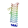
| ||||||||
|---|---|---|---|---|---|---|---|---|---|
| 1 |
| ||||||||
| Unit cell |
|
-Components
| #1: Protein | Mass: 27778.268 Da / Num. of mol.: 1 Source method: isolated from a genetically manipulated source Details: The protein adopts the repeated five residue (Rfr) fold comprised of a right-handed quadrilateral beta-helix Source: (gene. exp.) Nostoc sp. (bacteria) / Strain: PCC 7120 / Gene: HetL / Plasmid: pET-28B / Production host: |
|---|---|
| #2: Water | ChemComp-HOH /  Mass: 18.015 Da / Num. of mol.: 238 / Source method: isolated from a natural source / Formula: H2O Mass: 18.015 Da / Num. of mol.: 238 / Source method: isolated from a natural source / Formula: H2O |
-Experimental details
-Experiment
| Experiment | Method: X-RAY DIFFRACTION / Number of used crystals: 1 |
|---|
- Sample preparation
| Crystal | Density Matthews: 2.92 Å3/Da / Density % sol: 57.81 % |
|---|---|
| Crystal grow | Temperature: 295 K / Method: vapor diffusion, hanging drop / pH: 7.5 Details: 0.1M HEPES, 0.7M sodium phosphate monobasic, 0.7M potassium phosphate monobasic, 12.5% glycerol, protein concentration 7.5 mg/ml, VAPOR DIFFUSION, HANGING DROP, temperature 295K |
-Data collection
| Diffraction | Mean temperature: 100 K |
|---|---|
| Diffraction source | Source: ROTATING ANODE / Type: BRUKER AXS MICROSTAR / Wavelength: 1.54 Å |
| Detector | Type: BRUKER SMART 6000 / Detector: CCD / Date: Feb 13, 2008 / Details: mirrors |
| Radiation | Monochromator: graded thin film multilayer mirror Bragg Diffraction Protocol: SINGLE WAVELENGTH / Monochromatic (M) / Laue (L): M / Scattering type: x-ray |
| Radiation wavelength | Wavelength: 1.54 Å / Relative weight: 1 |
| Reflection | Resolution: 1.77→68.84 Å / Num. all: 32008 / Num. obs: 28767 / % possible obs: 89.9 % / Observed criterion σ(I): 3 / Redundancy: 38.58 % / Rsym value: 0.041 / Net I/σ(I): 61.22 |
| Reflection shell | Resolution: 1.77→1.87 Å / Redundancy: 38.58 % / Rmerge(I) obs: 0.3 / Mean I/σ(I) obs: 4.63 / Rsym value: 0.3 / % possible all: 58.3 |
- Processing
| Software |
| ||||||||||||||||||||||||||||||||||||||||||||||||||||||||||||||||||||||||||||||||||||||||||||||||||||||||||||||||||||||||||||||||||||||||||||||||||||||||||||||||||||||||||
|---|---|---|---|---|---|---|---|---|---|---|---|---|---|---|---|---|---|---|---|---|---|---|---|---|---|---|---|---|---|---|---|---|---|---|---|---|---|---|---|---|---|---|---|---|---|---|---|---|---|---|---|---|---|---|---|---|---|---|---|---|---|---|---|---|---|---|---|---|---|---|---|---|---|---|---|---|---|---|---|---|---|---|---|---|---|---|---|---|---|---|---|---|---|---|---|---|---|---|---|---|---|---|---|---|---|---|---|---|---|---|---|---|---|---|---|---|---|---|---|---|---|---|---|---|---|---|---|---|---|---|---|---|---|---|---|---|---|---|---|---|---|---|---|---|---|---|---|---|---|---|---|---|---|---|---|---|---|---|---|---|---|---|---|---|---|---|---|---|---|---|---|
| Refinement | Method to determine structure: sulfur anomalous scattering / Resolution: 2→28.41 Å / Cor.coef. Fo:Fc: 0.958 / Cor.coef. Fo:Fc free: 0.944 / SU B: 2.582 / SU ML: 0.075 / Cross valid method: THROUGHOUT / ESU R: 0.145 / ESU R Free: 0.131 / Stereochemistry target values: MAXIMUM LIKELIHOOD / Details: HYDROGENS HAVE BEEN ADDED IN THE RIDING POSITIONS
| ||||||||||||||||||||||||||||||||||||||||||||||||||||||||||||||||||||||||||||||||||||||||||||||||||||||||||||||||||||||||||||||||||||||||||||||||||||||||||||||||||||||||||
| Solvent computation | Ion probe radii: 0.8 Å / Shrinkage radii: 0.8 Å / VDW probe radii: 1.4 Å / Solvent model: MASK | ||||||||||||||||||||||||||||||||||||||||||||||||||||||||||||||||||||||||||||||||||||||||||||||||||||||||||||||||||||||||||||||||||||||||||||||||||||||||||||||||||||||||||
| Displacement parameters | Biso mean: 17.67 Å2
| ||||||||||||||||||||||||||||||||||||||||||||||||||||||||||||||||||||||||||||||||||||||||||||||||||||||||||||||||||||||||||||||||||||||||||||||||||||||||||||||||||||||||||
| Refinement step | Cycle: LAST / Resolution: 2→28.41 Å
| ||||||||||||||||||||||||||||||||||||||||||||||||||||||||||||||||||||||||||||||||||||||||||||||||||||||||||||||||||||||||||||||||||||||||||||||||||||||||||||||||||||||||||
| Refine LS restraints |
| ||||||||||||||||||||||||||||||||||||||||||||||||||||||||||||||||||||||||||||||||||||||||||||||||||||||||||||||||||||||||||||||||||||||||||||||||||||||||||||||||||||||||||
| LS refinement shell | Resolution: 2→2.052 Å / Total num. of bins used: 20
|
