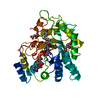
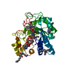
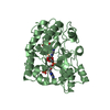
Delta4-3-oxosteroid 5beta-reductase / steroid dehydrogenase activity / bile acid catabolic process / C21-steroid hormone metabolic process / Delta4-3-oxosteroid 5beta-reductase activity / ketosteroid monooxygenase activity / Synthesis of bile acids and bile salts via 24-hydroxycholesterol / bile acid biosynthetic process / alcohol dehydrogenase (NADP+) activity / cholesterol catabolic process ...Delta4-3-oxosteroid 5beta-reductase / steroid dehydrogenase activity / bile acid catabolic process / C21-steroid hormone metabolic process / Delta4-3-oxosteroid 5beta-reductase activity / ketosteroid monooxygenase activity / Synthesis of bile acids and bile salts via 24-hydroxycholesterol / bile acid biosynthetic process / alcohol dehydrogenase (NADP+) activity / cholesterol catabolic process / aldose reductase (NADPH) activity / androgen metabolic process / Synthesis of bile acids and bile salts via 27-hydroxycholesterol / Synthesis of bile acids and bile salts via 7alpha-hydroxycholesterol / digestion / steroid binding / cytosol Similarity search - Function
Aldo-keto reductase family 1 member D1 / Aldo/keto reductase family putative active site signature. / Aldo/keto reductase family signature 1. / NADP-dependent oxidoreductase domain / Aldo/keto reductase family signature 2. / Aldo/keto reductase, conserved site / Aldo-keto reductase / NADP-dependent oxidoreductase domain / Aldo/keto reductase family / NADP-dependent oxidoreductase domain superfamily ...Aldo-keto reductase family 1 member D1 / Aldo/keto reductase family putative active site signature. / Aldo/keto reductase family signature 1. / NADP-dependent oxidoreductase domain / Aldo/keto reductase family signature 2. / Aldo/keto reductase, conserved site / Aldo-keto reductase / NADP-dependent oxidoreductase domain / Aldo/keto reductase family / NADP-dependent oxidoreductase domain superfamily / TIM Barrel / Alpha-Beta Barrel / Alpha Beta Similarity search - Domain/homology
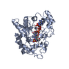
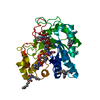
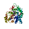
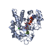
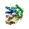
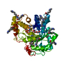
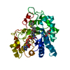
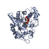
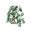
5-beta-DIHYDROTESTOSTERONE / NADP NICOTINAMIDE-ADENINE-DINUCLEOTIDE PHOSPHATE / Aldo-keto reductase family 1 member D1 Similarity search - Component
Biological species
Homo sapiens (human)
Method
X-RAY DIFFRACTION / FOURIER SYNTHESIS / Resolution: 2 Å
Mass: 18.015 Da / Num. of mol.: 588 / Source method: isolated from a natural source / Formula: H2O
Nonpolymer details
ON THE ACTIVE SITE OF MONOMER A AN INTERPRETABLE PEAK CORRESPONDING TO THE BINDING SITE OF 5-BETA- ...ON THE ACTIVE SITE OF MONOMER A AN INTERPRETABLE PEAK CORRESPONDING TO THE BINDING SITE OF 5-BETA-DIHYDROTESTOSTERONE WAS OBSERVED AND IT IS MISSING FROM THE MODEL.
-
Experimental details
-
Experiment
Experiment
Method: X-RAY DIFFRACTION
-
Sample preparation
Crystal
Density Matthews: 2.23 Å3/Da / Density % sol: 44.77 %
Crystal grow
Temperature: 277 K / Method: vapor diffusion, hanging drop / pH: 7 Details: ISOPROPANOL, PEG 4000, TRIS-HCL, PH 7.0, 10.0mM 5bDHT, VAPOR DIFFUSION, HANGING DROP, temperature 277K
-
Data collection
Diffraction
Mean temperature: 186 K
Diffraction source
Source: ROTATING ANODE / Type: RIGAKU / Wavelength: 1.5418 Å
In the structure databanks used in Yorodumi, some data are registered as the other names, "COVID-19 virus" and "2019-nCoV". Here are the details of the virus and the list of structure data.
Jan 31, 2019. EMDB accession codes are about to change! (news from PDBe EMDB page)
EMDB accession codes are about to change! (news from PDBe EMDB page)
The allocation of 4 digits for EMDB accession codes will soon come to an end. Whilst these codes will remain in use, new EMDB accession codes will include an additional digit and will expand incrementally as the available range of codes is exhausted. The current 4-digit format prefixed with “EMD-” (i.e. EMD-XXXX) will advance to a 5-digit format (i.e. EMD-XXXXX), and so on. It is currently estimated that the 4-digit codes will be depleted around Spring 2019, at which point the 5-digit format will come into force.
The EM Navigator/Yorodumi systems omit the EMD- prefix.
Related info.:Q: What is EMD? / ID/Accession-code notation in Yorodumi/EM Navigator
Yorodumi is a browser for structure data from EMDB, PDB, SASBDB, etc.
This page is also the successor to EM Navigator detail page, and also detail information page/front-end page for Omokage search.
The word "yorodu" (or yorozu) is an old Japanese word meaning "ten thousand". "mi" (miru) is to see.
Related info.:EMDB / PDB / SASBDB / Comparison of 3 databanks / Yorodumi Search / Aug 31, 2016. New EM Navigator & Yorodumi / Yorodumi Papers / Jmol/JSmol / Function and homology information / Changes in new EM Navigator and Yorodumi
 Movie
Movie Controller

 Yorodumi
 Open data
 Basic information
 Components
 Keywords
 Function and homology information
 Homo sapiens (human)
 X-RAY DIFFRACTION /
  Authors
 Citation
 Structure visualization
 Downloads & links
 Other downloads
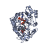

 PDBj
PDBj


 Assembly
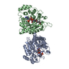


 Mass: 743.405 Da / Num. of mol.: 2 / Source method: obtained synthetically / Formula: C21H28N7O17P3
 Mass: 290.440 Da / Num. of mol.: 1 / Source method: obtained synthetically / Formula: C19H30O2
 Mass: 18.015 Da / Num. of mol.: 588 / Source method: isolated from a natural source / Formula: H2O
 Sample preparation
 Processing