 Movie
Movie Controller

[English] 日本語
 Yorodumi
- PDB-3dli: Crystal structure of a SAM dependent methyltransferase from Archa... -
+ Open data

- Basic information
| Entry | Database: PDB / ID: 3dli | ||||||
|---|---|---|---|---|---|---|---|
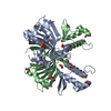
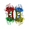
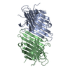
| Title | Crystal structure of a SAM dependent methyltransferase from Archaeoglobus fulgidus | ||||||
 Components Components | methyltransferase | ||||||
 Keywords Keywords | TRANSFERASE / 11116b / PSI-II / NYSGXRC / methyltransferase / Structural Genomics / Protein Structure Initiative / New York SGX Research Center for Structural Genomics | ||||||
| Function / homology | Methyltransferase domain / Vaccinia Virus protein VP39 / S-adenosyl-L-methionine-dependent methyltransferase superfamily / Rossmann fold / 3-Layer(aba) Sandwich / Alpha Beta / Methyltransferase domain-containing protein Function and homology information Function and homology information | ||||||
| Biological species |   Archaeoglobus fulgidus (archaea) Archaeoglobus fulgidus (archaea) | ||||||
| Method |  X-RAY DIFFRACTION / SYNCHROTRON / SAD / Resolution: 2.46 Å X-RAY DIFFRACTION / SYNCHROTRON / SAD / Resolution: 2.46 Å | ||||||
 Authors Authors | Agarwal, R. / Burley, S.K. / Swaminathan, S. / New York SGX Research Center for Structural Genomics (NYSGXRC) | ||||||
 Citation Citation | Journal: To be Published Title: Crystal structure of a SAM dependent methyltransferase from Archaeoglobus fulgidus Authors: Agarwal, R. / Burley, S.K. / Swaminathan, S. | ||||||
| History |
|
- Structure visualization
| Structure viewer | Molecule: MolmilJmol/JSmol |
|---|
- Downloads & links
-Download
| PDBx/mmCIF format | 3dli.cif.gz | 145.5 KB | Display | PDBx/mmCIF format |
|---|---|---|---|---|
| PDB format | pdb3dli.ent.gz | 115.4 KB | Display | PDB format |
| PDBx/mmJSON format | 3dli.json.gz | Tree view | PDBx/mmJSON format | |
| Others |  Other downloads Other downloads |
-Validation report
| Arichive directory | https://data.pdbj.org/pub/pdb/validation_reports/dl/3dliftp://data.pdbj.org/pub/pdb/validation_reports/dl/3dli | HTTPS FTP |
|---|
-Related structure data
| Similar structure data | |
|---|---|
| Other databases |


-Links
 PDBj
PDBj- Assembly
| Deposited unit | 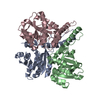
| ||||||||
|---|---|---|---|---|---|---|---|---|---|
| 1 |
| ||||||||
| Unit cell |
|
-Components
| #1: Protein | Mass: 28156.371 Da / Num. of mol.: 3 Source method: isolated from a genetically manipulated source Source: (gene. exp.) Archaeoglobus fulgidus (archaea) / Strain: DSM 4304 / Gene: AF_0046 / Plasmid: pSGX3(BC) / Production host:  #2: Water | ChemComp-HOH / |  Mass: 18.015 Da / Num. of mol.: 157 / Source method: isolated from a natural source / Formula: H2O Mass: 18.015 Da / Num. of mol.: 157 / Source method: isolated from a natural source / Formula: H2O |
|---|
-Experimental details
-Experiment
| Experiment | Method: X-RAY DIFFRACTION / Number of used crystals: 1 |
|---|
- Sample preparation
| Crystal | Density Matthews: 2.24 Å3/Da / Density % sol: 45 % |
|---|---|
| Crystal grow | Temperature: 298 K / Method: vapor diffusion, sitting drop / pH: 6.5 Details: 0.2M Ammonium sulfate, 0.1M Bis-tris, 25% PEG 3350, pH 6.5, VAPOR DIFFUSION, SITTING DROP, temperature 298K |
-Data collection
| Diffraction | Mean temperature: 100 K |
|---|---|
| Diffraction source | Source: SYNCHROTRON / Site: NSLS  / Beamline: X12C / Wavelength: 0.9792 Å / Beamline: X12C / Wavelength: 0.9792 Å |
| Detector | Type: ADSC QUANTUM 315 / Detector: CCD / Date: Jun 17, 2008 / Details: Mirrors |
| Radiation | Monochromator: SI-III / Protocol: SINGLE WAVELENGTH / Monochromatic (M) / Laue (L): M / Scattering type: x-ray |
| Radiation wavelength | Wavelength: 0.9792 Å / Relative weight: 1 |
| Reflection | Resolution: 2.46→50 Å / Num. all: 28155 / Num. obs: 28155 / % possible obs: 100 % / Observed criterion σ(I): 0 / Redundancy: 14.3 % / Biso Wilson estimate: 22.1 Å2 / Rmerge(I) obs: 0.08 / Net I/σ(I): 9.9 |
| Reflection shell | Resolution: 2.46→2.55 Å / Redundancy: 12.7 % / Rmerge(I) obs: 0.5 / Mean I/σ(I) obs: 2 / Num. unique all: 2757 / % possible all: 100 |
- Processing
| Software |
| ||||||||||||||||||||||||||||||
|---|---|---|---|---|---|---|---|---|---|---|---|---|---|---|---|---|---|---|---|---|---|---|---|---|---|---|---|---|---|---|---|
| Refinement | Method to determine structure: SAD Starting model: None Resolution: 2.46→41.12 Å / Rfactor Rfree error: 0.01 / Data cutoff high absF: 71812.77 / Data cutoff low absF: 0 / Isotropic thermal model: RESTRAINED / Cross valid method: THROUGHOUT / σ(F): 0 / Stereochemistry target values: Engh & Huber
| ||||||||||||||||||||||||||||||
| Solvent computation | Solvent model: FLAT MODEL / Bsol: 32.3972 Å2 / ksol: 0.375567 e/Å3 | ||||||||||||||||||||||||||||||
| Displacement parameters | Biso mean: 30.3 Å2
| ||||||||||||||||||||||||||||||
| Refine analyze |
| ||||||||||||||||||||||||||||||
| Refinement step | Cycle: LAST / Resolution: 2.46→41.12 Å
| ||||||||||||||||||||||||||||||
| Refine LS restraints |
| ||||||||||||||||||||||||||||||
| LS refinement shell | Resolution: 2.46→2.61 Å / Rfactor Rfree error: 0.027 / Total num. of bins used: 6
| ||||||||||||||||||||||||||||||
| Xplor file |
|
