 Movie
Movie Controller


+ Open data

- Basic information
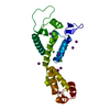
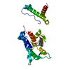
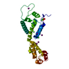
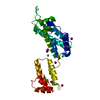
| Entry | Database: PDB / ID: 3dik | ||||||
|---|---|---|---|---|---|---|---|
| Title | Pseudo-atomic model of the HIV-1 CA hexameric lattice | ||||||
 Components Components | Capsid protein p24 | ||||||
 Keywords Keywords | VIRAL PROTEIN / MATURE RETROVIRAL CAPSID / FULLERENE CONE / HEXAMER / AIDS / Aspartyl protease / Capsid maturation / Capsid protein / DNA integration / DNA recombination / DNA-directed DNA polymerase / Endonuclease / Hydrolase / Lipoprotein / Magnesium / Metal-binding / Multifunctional enzyme / Myristate / Nuclease / Nucleotidyltransferase / Nucleus / Phosphoprotein / Protease / RNA-binding / RNA-directed DNA polymerase / Transferase / Viral nucleoprotein / Virion / Zinc-finger | ||||||
| Function / homology |  Function and homology information Function and homology informationHIV-1 retropepsin / symbiont-mediated activation of host apoptosis / retroviral ribonuclease H / exoribonuclease H / exoribonuclease H activity / DNA integration / viral genome integration into host DNA / establishment of integrated proviral latency / RNA-directed DNA polymerase / RNA stem-loop binding ...HIV-1 retropepsin / symbiont-mediated activation of host apoptosis / retroviral ribonuclease H / exoribonuclease H / exoribonuclease H activity / DNA integration / viral genome integration into host DNA / establishment of integrated proviral latency / RNA-directed DNA polymerase / RNA stem-loop binding / viral penetration into host nucleus / host multivesicular body / RNA-directed DNA polymerase activity / RNA-DNA hybrid ribonuclease activity / Transferases; Transferring phosphorus-containing groups; Nucleotidyltransferases / host cell / viral nucleocapsid / DNA recombination / DNA-directed DNA polymerase / aspartic-type endopeptidase activity / Hydrolases; Acting on ester bonds / DNA-directed DNA polymerase activity / symbiont-mediated suppression of host gene expression / viral translational frameshifting / symbiont entry into host cell / lipid binding / host cell nucleus / host cell plasma membrane / virion membrane / structural molecule activity / proteolysis / DNA binding / zinc ion binding Similarity search - Function | ||||||
| Biological species |   Human immunodeficiency virus type 1 Human immunodeficiency virus type 1 | ||||||
| Method | ELECTRON CRYSTALLOGRAPHY / electron crystallography / cryo EM / Resolution: 9 Å | ||||||
 Authors Authors | Ganser-Pornillos, B.K. / Cheng, A. / Yeager, M. | ||||||
 Citation Citation | Journal: Cell / Year: 2007 Title: Structure of full-length HIV-1 CA: a model for the mature capsid lattice. Authors: Barbie K Ganser-Pornillos / Anchi Cheng / Mark Yeager /  Abstract: The capsids of mature retroviruses perform the essential function of organizing the viral genome for efficient replication. These capsids are modeled as fullerene structures composed of closed ...The capsids of mature retroviruses perform the essential function of organizing the viral genome for efficient replication. These capsids are modeled as fullerene structures composed of closed hexameric arrays of the viral CA protein, but a high-resolution structure of the lattice has remained elusive. A three-dimensional map of two-dimensional crystals of the R18L mutant of HIV-1 CA was derived by electron cryocrystallography. The docking of high-resolution domain structures into the map yielded the first unambiguous model for full-length HIV-1 CA. Three important protein-protein assembly interfaces are required for capsid formation. Each CA hexamer is composed of an inner ring of six N-terminal domains and an outer ring of C-terminal domains that form dimeric linkers connecting neighboring hexamers. Interactions between the two domains of CA further stabilize the hexamer and provide a structural explanation for the mechanism of action of known HIV-1 assembly inhibitors. | ||||||
| History |
|
- Structure visualization
| Movie |
Movie viewer |
|---|---|
| Structure viewer | Molecule: MolmilJmol/JSmol |
- Downloads & links
-Download
| PDBx/mmCIF format | 3dik.cif.gz | 18 KB | Display | PDBx/mmCIF format |
|---|---|---|---|---|
| PDB format | pdb3dik.ent.gz | 7.5 KB | Display | PDB format |
| PDBx/mmJSON format | 3dik.json.gz | Tree view | PDBx/mmJSON format | |
| Others |  Other downloads Other downloads |
-Validation report
| Arichive directory | https://data.pdbj.org/pub/pdb/validation_reports/di/3dikftp://data.pdbj.org/pub/pdb/validation_reports/di/3dik | HTTPS FTP |
|---|
-Related structure data
| Related structure data |  1529MC M: map data used to model this data C: citing same article ( |
|---|---|
| Similar structure data |


-Links
 PDBj
PDBj
- Assembly
| Deposited unit | 
| ||||||||
|---|---|---|---|---|---|---|---|---|---|
| 1 |
| ||||||||
| Unit cell |
|
-Components
| #1: Protein | Mass: 24498.084 Da / Num. of mol.: 1 Source method: isolated from a genetically manipulated source Source: (gene. exp.) Human immunodeficiency virus type 1 / Gene: gag-pol / Plasmid: pET11a / Production host:  |
|---|
-Experimental details
-Experiment
| Experiment | Method: ELECTRON CRYSTALLOGRAPHY |
|---|---|
| EM experiment | Aggregation state: 2D ARRAY / 3D reconstruction method: electron crystallography |
- Sample preparation
| Component |
| ||||||||||||||||||||
|---|---|---|---|---|---|---|---|---|---|---|---|---|---|---|---|---|---|---|---|---|---|
| Buffer solution | Name: 17.5%(w/v) PEG 20,000, 50mM Na Cacodylate, 100mM Calcium Acetate pH: 6.5 Details: 17.5%(w/v) PEG 20,000, 50mM Na Cacodylate, 100mM Calcium Acetate | ||||||||||||||||||||
| Specimen | Conc.: 32 mg/ml / Embedding applied: NO / Shadowing applied: NO / Staining applied: NO / Vitrification applied: YES | ||||||||||||||||||||
| Specimen support | Details: Carbon coated molybdenum grids | ||||||||||||||||||||
| Vitrification | Cryogen name: ETHANE Details: Washed with 0.1M KCl, blotted briefly, and plunged into liquid ethane |
-Data collection
| Experimental equipment |  Model: Tecnai F20 / Image courtesy: FEI Company |
|---|---|
| Microscopy | Model: FEI TECNAI F20 |
| Electron gun | Electron source:  FIELD EMISSION GUN / Accelerating voltage: 120 kV / Illumination mode: OTHER FIELD EMISSION GUN / Accelerating voltage: 120 kV / Illumination mode: OTHER |
| Electron lens | Mode: BRIGHT FIELD / Nominal magnification: 52000 X / Nominal defocus max: 1500 nm / Nominal defocus min: 400 nm / Cs: 2 mm |
| Specimen holder | Temperature: 90 K / Tilt angle max: 40.3 ° / Tilt angle min: 0 ° |
| Image recording | Electron dose: 10 e/Å2 / Film or detector model: KODAK SO-163 FILM |
| Radiation | Protocol: SINGLE WAVELENGTH / Monochromatic (M) / Laue (L): M / Scattering type: electron |
| Radiation wavelength | Relative weight: 1 |
- Processing
| EM software |
| |||||||||||||||||||||
|---|---|---|---|---|---|---|---|---|---|---|---|---|---|---|---|---|---|---|---|---|---|---|
| 3D reconstruction | Method: 2D crystallography / Resolution: 9 Å Details: THIS MODDEL CONTAINS CA ATOMS ONLY. THE TWO DOMAINS UNP RESIDUES 133-280 AND UNP RESIDUES 281-351 WERE ALLOWED TO MOVE INDEPENDENTLY. THAT IS WHY THE CONNECTIVITY BETWEEN THEM IS NOT WELL- ...Details: THIS MODDEL CONTAINS CA ATOMS ONLY. THE TWO DOMAINS UNP RESIDUES 133-280 AND UNP RESIDUES 281-351 WERE ALLOWED TO MOVE INDEPENDENTLY. THAT IS WHY THE CONNECTIVITY BETWEEN THEM IS NOT WELL-PRESERVED. MRC AND CCP4 PROGRAMS WERE USED. Symmetry type: 2D CRYSTAL | |||||||||||||||||||||
| Atomic model building | Protocol: RIGID BODY FIT / Space: REAL / Details: REFINEMENT PROTOCOL--RIGID BODY | |||||||||||||||||||||
| Atomic model building |
| |||||||||||||||||||||
| Refinement step | Cycle: LAST
|


