Journal: Cell / Year: 2007 Title: Structure of full-length HIV-1 CA: a model for the mature capsid lattice. Authors: Barbie K Ganser-Pornillos / Anchi Cheng / Mark Yeager / Abstract: The capsids of mature retroviruses perform the essential function of organizing the viral genome for efficient replication. These capsids are modeled as fullerene structures composed of closed ...The capsids of mature retroviruses perform the essential function of organizing the viral genome for efficient replication. These capsids are modeled as fullerene structures composed of closed hexameric arrays of the viral CA protein, but a high-resolution structure of the lattice has remained elusive. A three-dimensional map of two-dimensional crystals of the R18L mutant of HIV-1 CA was derived by electron cryocrystallography. The docking of high-resolution domain structures into the map yielded the first unambiguous model for full-length HIV-1 CA. Three important protein-protein assembly interfaces are required for capsid formation. Each CA hexamer is composed of an inner ring of six N-terminal domains and an outer ring of C-terminal domains that form dimeric linkers connecting neighboring hexamers. Interactions between the two domains of CA further stabilize the hexamer and provide a structural explanation for the mechanism of action of known HIV-1 assembly inhibitors.
History
Deposition
Jun 24, 2008
-
Header (metadata) release
Jun 24, 2008
-
Map release
Apr 2, 2009
-
Update
Oct 24, 2012
-
Current status
Oct 24, 2012
Processing site: PDBe / Status: Released
-
Structure visualization
Movie
Surface view with section colored by density value
A: 92.7 Å / B: 92.7 Å / C: 110 Å α: 90 ° / β: 90 ° / γ: 120 °
CCP4 map header:
mode
Image stored as Reals
Å/pix. X/Y/Z
1.03
1.03
0.92436974789916
M x/y/z
90
90
119
origin x/y/z
0.000
0.000
0.000
length x/y/z
92.700
92.700
110.000
α/β/γ
90.000
90.000
120.000
start NX/NY/NZ
-75
-75
-74
NX/NY/NZ
150
150
150
MAP C/R/S
1
2
3
start NC/NR/NS
-90
-90
-60
NC/NR/NS
181
181
121
D min/max/mean
-241.730
250.000
-0.139
-
Supplemental data
-
Sample components
-
Entire : HIV-1 CA R18L
Entire
Name: HIV-1 CA R18L
Components
Sample: HIV-1 CA R18L
Protein or peptide: HIV-1 CA protein
-
Supramolecule #1000: HIV-1 CA R18L
Supramolecule
Name: HIV-1 CA R18L / type: sample / ID: 1000 / Oligomeric state: 6 / Number unique components: 1
Molecular weight
Experimental: 25 KDa / Theoretical: 25 KDa / Method: mass spectrometry of monomer
-
Macromolecule #1: HIV-1 CA protein
Macromolecule
Name: HIV-1 CA protein / type: protein_or_peptide / ID: 1 / Name.synonym: HIV-1 CA protein / Details: mass spectrometry of virus / Oligomeric state: hexamer / Recombinant expression: Yes
Source (natural)
Organism: Human immunodeficiency virus 1 / synonym: HIV-1
Molecular weight
Experimental: 25 KDa / Theoretical: 25 KDa
Recombinant expression
Organism: Escherichia coli (E. coli) / Recombinant plasmid: pET11a
Type: NEGATIVE Details: Grids were absorbed with crystals, washed with 0.1M KCl, blotted, and flash frozen in liquid ethane
Grid
Details: 300 mesh molybdemun
Vitrification
Cryogen name: ETHANE / Instrument: OTHER / Method: blot for 2 sec before plungin
Details
crstal grown by direct addition of PEG solution
Crystal formation
Details: crstal grown by direct addition of PEG solution
-
Electron microscopy
Microscope
FEI TECNAI F20
Alignment procedure
Legacy - Astigmatism: objective lens astigmatism was corrected at 100,000 times magnification
Image recording
Category: FILM / Film or detector model: KODAK SO-163 FILM / Digitization - Scanner: ZEISS SCAI / Digitization - Sampling interval: 7 µm / Number real images: 43 / Average electron dose: 15 e/Å2
Tilt angle min
0
Electron beam
Acceleration voltage: 120 kV / Electron source: FIELD EMISSION GUN
Electron optics
Illumination mode: OTHER / Imaging mode: BRIGHT FIELD / Cs: 2 mm / Nominal defocus max: 1.5 µm / Nominal defocus min: 0.4 µm / Nominal magnification: 50000
Sample stage
Specimen holder: Gatan 626 / Specimen holder model: GATAN LIQUID NITROGEN / Tilt angle max: 40 / Tilt series - Axis1 - Min angle: 0 ° / Tilt series - Axis1 - Max angle: 40 °
Experimental equipment
Model: Tecnai F20 / Image courtesy: FEI Company
-
Image processing
Final reconstruction
Resolution.type: BY AUTHOR / Resolution: 9.0 Å / Software - Name: MRC
Crystal parameters
Unit cell - A: 92.7 Å / Unit cell - B: 92.7 Å / Unit cell - C: 110 Å / Unit cell - γ: 120 ° / Unit cell - α: 90 ° / Unit cell - β: 90 ° / Plane group: P 6
In the structure databanks used in Yorodumi, some data are registered as the other names, "COVID-19 virus" and "2019-nCoV". Here are the details of the virus and the list of structure data.
Jan 31, 2019. EMDB accession codes are about to change! (news from PDBe EMDB page)
EMDB accession codes are about to change! (news from PDBe EMDB page)
The allocation of 4 digits for EMDB accession codes will soon come to an end. Whilst these codes will remain in use, new EMDB accession codes will include an additional digit and will expand incrementally as the available range of codes is exhausted. The current 4-digit format prefixed with “EMD-” (i.e. EMD-XXXX) will advance to a 5-digit format (i.e. EMD-XXXXX), and so on. It is currently estimated that the 4-digit codes will be depleted around Spring 2019, at which point the 5-digit format will come into force.
The EM Navigator/Yorodumi systems omit the EMD- prefix.
Related info.:Q: What is EMD? / ID/Accession-code notation in Yorodumi/EM Navigator
Yorodumi is a browser for structure data from EMDB, PDB, SASBDB, etc.
This page is also the successor to EM Navigator detail page, and also detail information page/front-end page for Omokage search.
The word "yorodu" (or yorozu) is an old Japanese word meaning "ten thousand". "mi" (miru) is to see.
Related info.:EMDB / PDB / SASBDB / Comparison of 3 databanks / Yorodumi Search / Aug 31, 2016. New EM Navigator & Yorodumi / Yorodumi Papers / Jmol/JSmol / Function and homology information / Changes in new EM Navigator and Yorodumi
 Movie
Movie Controller


 Open data
 Basic information
 Map data
 Sample
 Keywords
 Function and homology information
 Human immunodeficiency virus 1
 Authors
 Citation
 Structure visualization
 Downloads & links
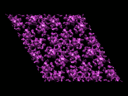 1529.gif
 http://ftp.pdbj.org/pub/emdb/structures/EMD-1529

 Z (Sec.)
 Y (Row.)
 X (Col.)


 Sample components
 Processing
 Electron microscopy
 FIELD EMISSION GUN


