 Movie
Movie Controller

[English] 日本語
 Yorodumi
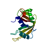
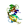
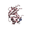
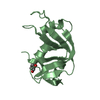
- PDB-3dib: Crystal structure of bovine pancreatic ribonuclease A variant (I106A) -
+ Open data

- Basic information
| Entry | Database: PDB / ID: 3dib | ||||||
|---|---|---|---|---|---|---|---|
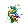
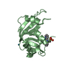
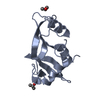
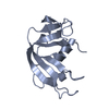
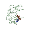
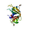
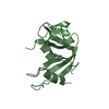
| Title | Crystal structure of bovine pancreatic ribonuclease A variant (I106A) | ||||||
 Components Components | Ribonuclease pancreatic | ||||||
 Keywords Keywords | HYDROLASE / ribonuclease / RNase A / Bovine pancreas / Endonuclease / Glycation / Glycoprotein / Nuclease / Secreted | ||||||
| Function / homology |  Function and homology information Function and homology informationpancreatic ribonuclease / ribonuclease A activity / RNA nuclease activity / nucleic acid binding / defense response to Gram-positive bacterium / lyase activity / extracellular region Similarity search - Function | ||||||
| Biological species |  | ||||||
| Method |  X-RAY DIFFRACTION / MOLECULAR REPLACEMENT / Resolution: 1.4 Å X-RAY DIFFRACTION / MOLECULAR REPLACEMENT / Resolution: 1.4 Å | ||||||
 Authors Authors | Kurpiewska, K. / Font, J. / Ribo, M. / Vilanova, M. / Lewinski, K. | ||||||
 Citation Citation | Journal: Proteins / Year: 2009 Title: X-ray crystallographic studies of RNase A variants engineered at the most destabilizing positions of the main hydrophobic core: further insight into protein stability Authors: Kurpiewska, K. / Font, J. / Ribo, M. / Vilanova, M. / Lewinski, K. | ||||||
| History |
|
- Structure visualization
| Structure viewer | Molecule: MolmilJmol/JSmol |
|---|
- Downloads & links
-Download
| PDBx/mmCIF format | 3dib.cif.gz | 43.3 KB | Display | PDBx/mmCIF format |
|---|---|---|---|---|
| PDB format | pdb3dib.ent.gz | 29.3 KB | Display | PDB format |
| PDBx/mmJSON format | 3dib.json.gz | Tree view | PDBx/mmJSON format | |
| Others |  Other downloads Other downloads |
-Validation report
| Summary document | 3dib_validation.pdf.gz | 436.2 KB | Display | wwPDB validaton report |
|---|---|---|---|---|
| Full document | 3dib_full_validation.pdf.gz | 438.8 KB | Display | |
| Data in XML | 3dib_validation.xml.gz | 10.4 KB | Display | |
| Data in CIF | 3dib_validation.cif.gz | 15.1 KB | Display | |
| Arichive directory | https://data.pdbj.org/pub/pdb/validation_reports/di/3dibftp://data.pdbj.org/pub/pdb/validation_reports/di/3dib | HTTPS FTP |
-Related structure data
| Related structure data |  3dh5C 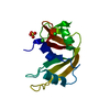 3dh6C 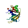 3di7C 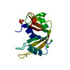 3di8C 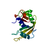 3di9C 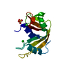 3dicC 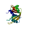 1fs3S C: citing same article ( S: Starting model for refinement |
|---|---|
| Similar structure data |


-Links
 PDBj
PDBj

- Assembly
| Deposited unit | 
| ||||||||
|---|---|---|---|---|---|---|---|---|---|
| 1 |
| ||||||||
| 2 | 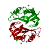
| ||||||||
| Unit cell |
| ||||||||
| Components on special symmetry positions |
|
-Components
| #1: Protein | Mass: 13666.246 Da / Num. of mol.: 1 / Mutation: I106A Source method: isolated from a genetically manipulated source Source: (gene. exp.)  | ||||||
|---|---|---|---|---|---|---|---|
| #2: Chemical | ChemComp-CL /   Mass: 35.453 Da / Num. of mol.: 5 / Source method: obtained synthetically / Formula: Cl Mass: 35.453 Da / Num. of mol.: 5 / Source method: obtained synthetically / Formula: Cl#3: Chemical | ChemComp-SO4 / |   Mass: 96.063 Da / Num. of mol.: 1 / Source method: obtained synthetically / Formula: SO4 Mass: 96.063 Da / Num. of mol.: 1 / Source method: obtained synthetically / Formula: SO4#4: Water | ChemComp-HOH / |  Mass: 18.015 Da / Num. of mol.: 239 / Source method: isolated from a natural source / Formula: H2O Mass: 18.015 Da / Num. of mol.: 239 / Source method: isolated from a natural source / Formula: H2OHas protein modification | Y | |
-Experimental details
-Experiment
| Experiment | Method: X-RAY DIFFRACTION / Number of used crystals: 1 |
|---|
- Sample preparation
| Crystal | Density Matthews: 2.79 Å3/Da / Density % sol: 55.98 % |
|---|---|
| Crystal grow | Temperature: 298 K / Method: vapor diffusion, hanging drop / pH: 6 Details: ammonium sulfate, sodium chloride, sodium acetate, pH 6.0, vapor diffusion, hanging drop, temperature 298K |
-Data collection
| Diffraction | Mean temperature: 273 K |
|---|---|
| Diffraction source | Source: SEALED TUBE / Type: ENRAF-NONIUS FR590 / Wavelength: 0.7107 Å |
| Detector | Type: Nonius Kappa CCD / Detector: CCD / Date: Aug 25, 2005 |
| Radiation | Monochromator: GRAPHITE / Protocol: SINGLE WAVELENGTH / Monochromatic (M) / Laue (L): M / Scattering type: x-ray |
| Radiation wavelength | Wavelength: 0.7107 Å / Relative weight: 1 |
| Reflection | Resolution: 1.4→10 Å / Num. obs: 34686 / Biso Wilson estimate: 7.9 Å2 |
- Processing
| Software |
| ||||||||||||||||||||||||||||||||||||||||||||||||||||||||||||||||||||||||||||||||
|---|---|---|---|---|---|---|---|---|---|---|---|---|---|---|---|---|---|---|---|---|---|---|---|---|---|---|---|---|---|---|---|---|---|---|---|---|---|---|---|---|---|---|---|---|---|---|---|---|---|---|---|---|---|---|---|---|---|---|---|---|---|---|---|---|---|---|---|---|---|---|---|---|---|---|---|---|---|---|---|---|---|
| Refinement | Method to determine structure: MOLECULAR REPLACEMENT Starting model: PDB ENTRY 1FS3 Resolution: 1.4→10 Å / Rfactor Rfree error: 0.005 / Data cutoff high absF: 154426.12 / Data cutoff low absF: 0 / Isotropic thermal model: RESTRAINED / Cross valid method: THROUGHOUT / σ(F): 0
| ||||||||||||||||||||||||||||||||||||||||||||||||||||||||||||||||||||||||||||||||
| Solvent computation | Solvent model: FLAT MODEL / Bsol: 39.3828 Å2 / ksol: 0.467979 e/Å3 | ||||||||||||||||||||||||||||||||||||||||||||||||||||||||||||||||||||||||||||||||
| Displacement parameters | Biso mean: 12.6 Å2
| ||||||||||||||||||||||||||||||||||||||||||||||||||||||||||||||||||||||||||||||||
| Refine analyze |
| ||||||||||||||||||||||||||||||||||||||||||||||||||||||||||||||||||||||||||||||||
| Refinement step | Cycle: LAST / Resolution: 1.4→10 Å
| ||||||||||||||||||||||||||||||||||||||||||||||||||||||||||||||||||||||||||||||||
| Refine LS restraints |
| ||||||||||||||||||||||||||||||||||||||||||||||||||||||||||||||||||||||||||||||||
| LS refinement shell | Resolution: 1.4→1.49 Å / Rfactor Rfree error: 0.014 / Total num. of bins used: 6
| ||||||||||||||||||||||||||||||||||||||||||||||||||||||||||||||||||||||||||||||||
| Xplor file |
|
