 Movie
Movie Controller

[English] 日本語
 Yorodumi
- PDB-3dha: An Ultral High Resolution Structure of N-Acyl Homoserine Lactone ... -
+ Open data

- Basic information
| Entry | Database: PDB / ID: 3dha | ||||||
|---|---|---|---|---|---|---|---|
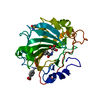
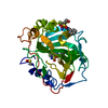
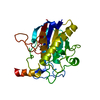
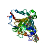
| Title | An Ultral High Resolution Structure of N-Acyl Homoserine Lactone Hydrolase with the Product N-Hexanoyl-L-Homoserine Bound at An Alternative Site | ||||||
 Components Components | N-Acyl Homoserine Lactone Hydrolase | ||||||
 Keywords Keywords | HYDROLASE / Zinc Bimetallohydrolase / Quorum Quenching / N-Acyl Homoserine lactone / Alternative Binding Site / Product Complex / AHL Lactonase | ||||||
| Function / homology |  Function and homology information Function and homology informationlactone catabolic process / quorum-quenching N-acyl-homoserine lactonase / acyl-L-homoserine-lactone lactonohydrolase activity / metal ion binding Similarity search - Function | ||||||
| Biological species |  | ||||||
| Method |  X-RAY DIFFRACTION / SYNCHROTRON / MOLECULAR REPLACEMENT / Resolution: 0.95 Å X-RAY DIFFRACTION / SYNCHROTRON / MOLECULAR REPLACEMENT / Resolution: 0.95 Å | ||||||
 Authors Authors | Liu, D. / Momb, J. / Thomas, P.W. / Moulin, A. / Petsko, G.A. / Fast, W. / Ringe, D. | ||||||
 Citation Citation | Journal: Biochemistry / Year: 2008 Title: Mechanism of the quorum-quenching lactonase (AiiA) from Bacillus thuringiensis. 1. Product-bound structures. Authors: Liu, D. / Momb, J. / Thomas, P.W. / Moulin, A. / Petsko, G.A. / Fast, W. / Ringe, D. #1: Journal: To be PublishedTitle: Mechanism of the Quorum-quenching Lactonase (AiiA) from Bacillus thuringensis: 2. Substrate Modeling and Active Site Mutations Authors: Momb, J. / Wang, C. / Liu, D. / Thomas, P.W. / Petsko, G.A. / Guo, H. / Ringe, D. / Fast, W. | ||||||
| History |
|
- Structure visualization
| Structure viewer | Molecule: MolmilJmol/JSmol |
|---|
- Downloads & links
-Download
| PDBx/mmCIF format | 3dha.cif.gz | 133.6 KB | Display | PDBx/mmCIF format |
|---|---|---|---|---|
| PDB format | pdb3dha.ent.gz | 102 KB | Display | PDB format |
| PDBx/mmJSON format | 3dha.json.gz | Tree view | PDBx/mmJSON format | |
| Others |  Other downloads Other downloads |
-Validation report
| Arichive directory | https://data.pdbj.org/pub/pdb/validation_reports/dh/3dhaftp://data.pdbj.org/pub/pdb/validation_reports/dh/3dha | HTTPS FTP |
|---|
-Related structure data
| Related structure data |  3dhbC  3dhcC  2a7mS S: Starting model for refinement C: citing same article ( |
|---|---|
| Similar structure data |


-Links
 PDBj
PDBj

- Assembly
| Deposited unit | 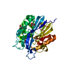
| ||||||||
|---|---|---|---|---|---|---|---|---|---|
| 1 |
| ||||||||
| Unit cell |
|
-Components
| #1: Protein | Mass: 28664.643 Da / Num. of mol.: 1 Source method: isolated from a genetically manipulated source Source: (gene. exp.) Description: The fusion protein, maltose binding protein, was cut off before crystallization using TEV protease resulting in 4 extra residues at the N terminus. Gene: aiiA / Plasmid: pMAL / Production host: References: UniProt: Q7B8B9, UniProt: P0CJ63*PLUS, Hydrolases; Acting on ester bonds; Carboxylic-ester hydrolases | ||||||
|---|---|---|---|---|---|---|---|
| #2: Chemical |   Mass: 65.409 Da / Num. of mol.: 2 / Source method: obtained synthetically / Formula: Zn Mass: 65.409 Da / Num. of mol.: 2 / Source method: obtained synthetically / Formula: Zn#3: Chemical | ChemComp-C6L / |   Mass: 217.262 Da / Num. of mol.: 1 / Source method: obtained synthetically / Formula: C10H19NO4 Mass: 217.262 Da / Num. of mol.: 1 / Source method: obtained synthetically / Formula: C10H19NO4#4: Chemical | ChemComp-GOL /   Mass: 92.094 Da / Num. of mol.: 4 / Source method: obtained synthetically / Formula: C3H8O3 Mass: 92.094 Da / Num. of mol.: 4 / Source method: obtained synthetically / Formula: C3H8O3#5: Water | ChemComp-HOH / |  Mass: 18.015 Da / Num. of mol.: 255 / Source method: isolated from a natural source / Formula: H2O Mass: 18.015 Da / Num. of mol.: 255 / Source method: isolated from a natural source / Formula: H2O |
-Experimental details
-Experiment
| Experiment | Method: X-RAY DIFFRACTION / Number of used crystals: 1 |
|---|
- Sample preparation
| Crystal | Density Matthews: 2.1 Å3/Da / Density % sol: 41.33 % |
|---|---|
| Crystal grow | Temperature: 298 K / Method: vapor diffusion, hanging drop / pH: 8.5 Details: Glycerol, Tris-HCl, PEG4000, MgCl2, N-hexanoyl-L-homoserine lactone, pH 8.5, VAPOR DIFFUSION, HANGING DROP, temperature 298K |
-Data collection
| Diffraction | Mean temperature: 100 K |
|---|---|
| Diffraction source | Source: SYNCHROTRON / Site: APS  / Beamline: 23-ID-D / Wavelength: 0.9184 Å / Beamline: 23-ID-D / Wavelength: 0.9184 Å |
| Detector | Type: MARMOSAIC 300 mm CCD / Detector: CCD / Date: Apr 20, 2006 / Details: K-B pair of biomorph mirrors |
| Radiation | Monochromator: double crystal monochromator / Protocol: SINGLE WAVELENGTH / Monochromatic (M) / Laue (L): M / Scattering type: x-ray |
| Radiation wavelength | Wavelength: 0.9184 Å / Relative weight: 1 |
| Reflection | Resolution: 0.95→50 Å / Num. obs: 141032 / % possible obs: 93.9 % / Observed criterion σ(F): 0 / Observed criterion σ(I): 0 / Redundancy: 6.3 % / Biso Wilson estimate: 15.9 Å2 / Rmerge(I) obs: 0.091 / Net I/σ(I): 12.5 |
| Reflection shell | Resolution: 0.95→0.98 Å / Redundancy: 2.2 % / Rmerge(I) obs: 0.371 / Mean I/σ(I) obs: 2 / Num. unique all: 9081 / % possible all: 61.9 |
- Processing
| Software |
| |||||||||||||||||||||||||||||||||
|---|---|---|---|---|---|---|---|---|---|---|---|---|---|---|---|---|---|---|---|---|---|---|---|---|---|---|---|---|---|---|---|---|---|---|
| Refinement | Method to determine structure: MOLECULAR REPLACEMENT Starting model: PDB entry 2A7M Resolution: 0.95→45 Å / Num. parameters: 22638 / Num. restraintsaints: 28771 / Isotropic thermal model: anisotropic / Cross valid method: FREE R / σ(F): 0 / σ(I): 2 / Stereochemistry target values: ENGH AND HUBER Details: ANISOTROPIC REFINEMENT REDUCED FREE R (NO CUTOFF) BY 0.0565
| |||||||||||||||||||||||||||||||||
| Displacement parameters | Biso mean: 21.8 Å2 | |||||||||||||||||||||||||||||||||
| Refine analyze | Luzzati coordinate error obs: 0.037 Å / Num. disordered residues: 18 / Occupancy sum hydrogen: 1951 / Occupancy sum non hydrogen: 2406.5 | |||||||||||||||||||||||||||||||||
| Refinement step | Cycle: LAST / Resolution: 0.95→45 Å
| |||||||||||||||||||||||||||||||||
| Refine LS restraints |
| |||||||||||||||||||||||||||||||||
| LS refinement shell | Resolution: 0.95→0.98 Å /
|
