Resolution: 1.6→1.686 Å / Redundancy: 7 % / Rmerge(I) obs: 0.87 / Mean I/σ(I) obs: 2.4 / Num. unique all: 3603 / Rsym value: 0.87 / % possible all: 80.49
-
Processing
Software
Name
Version
Classification
REFMAC
5.2.0005
refinement
Blu-Ice
datacollection
SCALEPACK
datascaling
SOLVE
phasing
Refinement
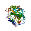
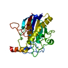
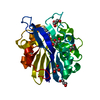
Method to determine structure: SAD / Resolution: 1.6→45.64 Å / Cor.coef. Fo:Fc: 0.967 / Cor.coef. Fo:Fc free: 0.948 / SU B: 4.105 / SU ML: 0.071 / Cross valid method: THROUGHOUT / σ(F): 0 / σ(I): 0 / ESU R: 0.099 / ESU R Free: 0.101 / Stereochemistry target values: MAXIMUM LIKELIHOOD Details: Atoms with little to no electron density are represented in this PDB file as having zero occupancy, and the atomic displacement parameters for these atoms have been set to zero. These atoms ...Details: Atoms with little to no electron density are represented in this PDB file as having zero occupancy, and the atomic displacement parameters for these atoms have been set to zero. These atoms have been included in this file with geometry corresponding to that which would be obtained in refinement, however they were not included in the calculations for the residuals. Residues ASP A 50 and ILE A 190 exist in generously allowed and disallowed sectors of the Ramachandran plot and exhibit outstanding omit map electron density. ILE A 142 is on the edge of the additionally allowed and generously allowed sector of the plot as calculated with PROCHECK and MOLEMAN2.
Rfactor
Num. reflection
% reflection
Selection details
Rfree
0.21068
1420
4.7 %
RANDOM
Rwork
0.16976
-
-
-
all
0.17171
28550
-
-
obs
0.17171
28550
91.22 %
-
Solvent computation
Ion probe radii: 0.8 Å / Shrinkage radii: 0.8 Å / VDW probe radii: 1.4 Å / Solvent model: BABINET MODEL WITH MASK
Displacement parameters
Biso mean: 26.651 Å2
Baniso -1
Baniso -2
Baniso -3
1-
0.85 Å2
0 Å2
0 Å2
2-
-
-0.96 Å2
0 Å2
3-
-
-
0.11 Å2
Refine analyze
Free
Obs
Luzzati coordinate error
0.32 Å
0.29 Å
Luzzati d res low
-
1.6 Å
Luzzati sigma a
0.17 Å
0.2 Å
Refinement step
Cycle: LAST / Resolution: 1.6→45.64 Å
Protein
Nucleic acid
Ligand
Solvent
Total
Num. atoms
1989
0
50
181
2220
Refine LS restraints
Refine-ID
Type
Dev ideal
Dev ideal target
Number
X-RAY DIFFRACTION
r_bond_refined_d
0.009
0.022
2173
X-RAY DIFFRACTION
r_angle_refined_deg
1.305
2.011
2931
X-RAY DIFFRACTION
r_dihedral_angle_1_deg
6.189
5
250
X-RAY DIFFRACTION
r_dihedral_angle_2_deg
31.409
25.368
95
X-RAY DIFFRACTION
r_dihedral_angle_3_deg
14.294
15
397
X-RAY DIFFRACTION
r_dihedral_angle_4_deg
19.213
15
6
X-RAY DIFFRACTION
r_chiral_restr
0.085
0.2
339
X-RAY DIFFRACTION
r_gen_planes_refined
0.005
0.02
1555
X-RAY DIFFRACTION
r_nbd_refined
0.201
0.2
1074
X-RAY DIFFRACTION
r_nbtor_refined
0.31
0.2
1481
X-RAY DIFFRACTION
r_xyhbond_nbd_refined
0.114
0.2
169
X-RAY DIFFRACTION
r_metal_ion_refined
0.033
0.2
3
X-RAY DIFFRACTION
r_symmetry_vdw_refined
0.204
0.2
60
X-RAY DIFFRACTION
r_symmetry_hbond_refined
0.102
0.2
17
X-RAY DIFFRACTION
r_mcbond_it
1.734
2
1317
X-RAY DIFFRACTION
r_mcangle_it
2.29
3
2098
X-RAY DIFFRACTION
r_scbond_it
3.712
4
957
X-RAY DIFFRACTION
r_scangle_it
5.173
6
833
LS refinement shell
Resolution: 1.6→1.686 Å / Total num. of bins used: 10
Rfactor
Num. reflection
% reflection
Rfree
0.299
192
-
Rwork
0.234
3603
-
obs
-
-
80.49 %
Refinement TLS params.
Method: refined / Origin x: 19.919 Å / Origin y: 30.759 Å / Origin z: 29.698 Å
11
12
13
21
22
23
31
32
33
T
-0.0668 Å2
0.0156 Å2
0.0107 Å2
-
-0.0607 Å2
-0.0141 Å2
-
-
-0.0895 Å2
L
1.8182 °2
-0.5265 °2
0.241 °2
-
2.2137 °2
0.1824 °2
-
-
0.9932 °2
S
-0.0854 Å °
-0.2562 Å °
0.0257 Å °
0.1552 Å °
0.0478 Å °
0.148 Å °
0.0047 Å °
-0.1194 Å °
0.0375 Å °
+
About Yorodumi
-
News
-
Feb 9, 2022. New format data for meta-information of EMDB entries
New format data for meta-information of EMDB entries
Version 3 of the EMDB header file is now the official format.
The previous official version 1.9 will be removed from the archive.
In the structure databanks used in Yorodumi, some data are registered as the other names, "COVID-19 virus" and "2019-nCoV". Here are the details of the virus and the list of structure data.
Jan 31, 2019. EMDB accession codes are about to change! (news from PDBe EMDB page)
EMDB accession codes are about to change! (news from PDBe EMDB page)
The allocation of 4 digits for EMDB accession codes will soon come to an end. Whilst these codes will remain in use, new EMDB accession codes will include an additional digit and will expand incrementally as the available range of codes is exhausted. The current 4-digit format prefixed with “EMD-” (i.e. EMD-XXXX) will advance to a 5-digit format (i.e. EMD-XXXXX), and so on. It is currently estimated that the 4-digit codes will be depleted around Spring 2019, at which point the 5-digit format will come into force.
The EM Navigator/Yorodumi systems omit the EMD- prefix.
Related info.:Q: What is EMD? / ID/Accession-code notation in Yorodumi/EM Navigator
Yorodumi is a browser for structure data from EMDB, PDB, SASBDB, etc.
This page is also the successor to EM Navigator detail page, and also detail information page/front-end page for Omokage search.
The word "yorodu" (or yorozu) is an old Japanese word meaning "ten thousand". "mi" (miru) is to see.
Related info.:EMDB / PDB / SASBDB / Comparison of 3 databanks / Yorodumi Search / Aug 31, 2016. New EM Navigator & Yorodumi / Yorodumi Papers / Jmol/JSmol / Function and homology information / Changes in new EM Navigator and Yorodumi
 Movie
Movie Controller

 Yorodumi
 Open data
 Basic information
 Components
 Keywords
 Function and homology information
 X-RAY DIFFRACTION /
  Authors
 Citation
 Structure visualization
 Downloads & links
 Other downloads
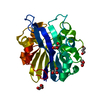

 PDBj
PDBj

 Assembly

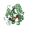
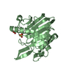
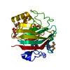
 Mass: 65.409 Da / Num. of mol.: 2 / Source method: obtained synthetically / Formula: Zn
 Mass: 92.094 Da / Num. of mol.: 8 / Source method: obtained synthetically / Formula: C3H8O3
 Mass: 18.015 Da / Num. of mol.: 181 / Source method: isolated from a natural source / Formula: H2O
 Sample preparation
 Processing