 Movie
Movie Controller

[English] 日本語
 Yorodumi
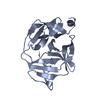
- PDB-3dex: Crystal structure of SAV_2001 protein from Streptomyces avermitil... -
+ Open data

- Basic information
| Entry | Database: PDB / ID: 3dex | ||||||
|---|---|---|---|---|---|---|---|
| Title | Crystal structure of SAV_2001 protein from Streptomyces avermitilis, Northeast Structural Genomics Consortium Target SvR107. | ||||||
 Components Components | SAV_2001 | ||||||
 Keywords Keywords | structural genomics / unknown function / alpha-beta protein / PSI-2 / Protein Structure Initiative / Northeast Structural Genomics Consortium / NESG | ||||||
| Function / homology | Selenoprotein, Rdx-type / Rdx family / Glutaredoxin / Glutaredoxin / Thioredoxin-like superfamily / 3-Layer(aba) Sandwich / Alpha Beta / Selenoprotein W-related protein Function and homology information Function and homology information | ||||||
| Biological species |  Streptomyces avermitilis (bacteria) Streptomyces avermitilis (bacteria) | ||||||
| Method |  X-RAY DIFFRACTION / SYNCHROTRON / MOLECULAR REPLACEMENT / Resolution: 2.7 Å X-RAY DIFFRACTION / SYNCHROTRON / MOLECULAR REPLACEMENT / Resolution: 2.7 Å | ||||||
 Authors Authors | Forouhar, F. / Neely, H. / Seetharaman, J. / Janjua, H. / Fang, Y. / Xiao, R. / Cunningham, K. / Ma, L.-C. / Owen, L.A. / Chen, C.X. ...Forouhar, F. / Neely, H. / Seetharaman, J. / Janjua, H. / Fang, Y. / Xiao, R. / Cunningham, K. / Ma, L.-C. / Owen, L.A. / Chen, C.X. / Acton, T.B. / Montelione, G.T. / Hunt, J.F. / Tong, L. / Northeast Structural Genomics Consortium (NESG) | ||||||
 Citation Citation | Journal: To be Published Title: Crystal structure of SAV_2001 protein from Streptomyces avermitilis, Northeast Structural Genomics Consortium Target SvR107. Authors: Forouhar, F. / Neely, H. / Seetharaman, J. / Janjua, H. / Fang, Y. / Xiao, R. / Cunningham, K. / Ma, L.-C. / Owen, L.A. / Chen, C.X. / Acton, T.B. / Montelione, G.T. / Hunt, J.F. / Tong, L. | ||||||
| History |
|
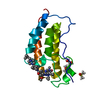
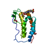
- Structure visualization
| Structure viewer | Molecule: MolmilJmol/JSmol |
|---|
- Downloads & links
-Download
| PDBx/mmCIF format | 3dex.cif.gz | 136.3 KB | Display | PDBx/mmCIF format |
|---|---|---|---|---|
| PDB format | pdb3dex.ent.gz | 110 KB | Display | PDB format |
| PDBx/mmJSON format | 3dex.json.gz | Tree view | PDBx/mmJSON format | |
| Others |  Other downloads Other downloads |
-Validation report
| Arichive directory | https://data.pdbj.org/pub/pdb/validation_reports/de/3dexftp://data.pdbj.org/pub/pdb/validation_reports/de/3dex | HTTPS FTP |
|---|
-Related structure data
| Related structure data | 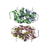 2okaS S: Starting model for refinement |
|---|---|
| Similar structure data | |
| Other databases |


-Links
 PDBj
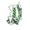
PDBj- Assembly
| Deposited unit | 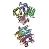
| ||||||||
|---|---|---|---|---|---|---|---|---|---|
| 1 | 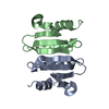
| ||||||||
| 2 | 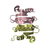
| ||||||||
| 3 | 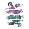
| ||||||||
| 4 | 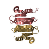
| ||||||||
| Unit cell |
| ||||||||
| Details | authors state that the biological assembly is possibly dimer. |
-Components
| #1: Protein | Mass: 12443.153 Da / Num. of mol.: 8 Source method: isolated from a genetically manipulated source Source: (gene. exp.) Streptomyces avermitilis (bacteria) / Strain: MA-4680 / Gene: SAV2001, SAV_2001 / Plasmid: BL21 / Production host: Has protein modification | Y | |
|---|
-Experimental details
-Experiment
| Experiment | Method: X-RAY DIFFRACTION / Number of used crystals: 1 |
|---|
- Sample preparation
| Crystal | Density Matthews: 1.84 Å3/Da / Density % sol: 33.16 % |
|---|---|
| Crystal grow | Temperature: 291 K / Method: vapor diffusion, hanging drop / pH: 7.5 Details: Protein solution: 10 mM Tris (pH 7.5), 100 mM sodium chloride, and 5 mM DTT. Reservoir solution: 14% PEG 3350, 0.15 M K/Na-tartrate, and 10% glycerol., VAPOR DIFFUSION, HANGING DROP, temperature 291K |
-Data collection
| Diffraction | Mean temperature: 100 K |
|---|---|
| Diffraction source | Source: SYNCHROTRON / Site: NSLS  / Beamline: X4C / Wavelength: 0.979 Å / Beamline: X4C / Wavelength: 0.979 Å |
| Detector | Type: MAR CCD 165 mm / Detector: CCD / Details: mirrors. |
| Radiation | Monochromator: Si 111 CHANNEL / Protocol: SINGLE WAVELENGTH / Monochromatic (M) / Laue (L): M / Scattering type: x-ray |
| Radiation wavelength | Wavelength: 0.979 Å / Relative weight: 1 |
| Reflection | Resolution: 2.7→30 Å / Num. all: 41024 / Num. obs: 39958 / % possible obs: 97.4 % / Observed criterion σ(F): 0 / Observed criterion σ(I): 0 / Redundancy: 3.5 % / Biso Wilson estimate: 34.2 Å2 / Rmerge(I) obs: 0.118 / Rsym value: 0.101 / Net I/σ(I): 12.42 |
| Reflection shell | Resolution: 2.7→2.8 Å / Redundancy: 3.4 % / Rmerge(I) obs: 0.472 / Mean I/σ(I) obs: 2.58 / Rsym value: 0.524 / % possible all: 94.7 |
- Processing
| Software |
| ||||||||||||||||||||||||||||||||||||||||||||||||||||||||||||
|---|---|---|---|---|---|---|---|---|---|---|---|---|---|---|---|---|---|---|---|---|---|---|---|---|---|---|---|---|---|---|---|---|---|---|---|---|---|---|---|---|---|---|---|---|---|---|---|---|---|---|---|---|---|---|---|---|---|---|---|---|---|
| Refinement | Method to determine structure: MOLECULAR REPLACEMENT Starting model: 2OKA Resolution: 2.7→19.99 Å / Rfactor Rfree error: 0.005 / Data cutoff high absF: 567738.9 / Data cutoff low absF: 0 / Isotropic thermal model: OVERALL / Cross valid method: THROUGHOUT / σ(F): 2 / Stereochemistry target values: Engh & Huber
| ||||||||||||||||||||||||||||||||||||||||||||||||||||||||||||
| Solvent computation | Solvent model: FLAT MODEL / Bsol: 34.9882 Å2 / ksol: 0.4 e/Å3 | ||||||||||||||||||||||||||||||||||||||||||||||||||||||||||||
| Displacement parameters | Biso mean: 34.1 Å2
| ||||||||||||||||||||||||||||||||||||||||||||||||||||||||||||
| Refine analyze |
| ||||||||||||||||||||||||||||||||||||||||||||||||||||||||||||
| Refinement step | Cycle: LAST / Resolution: 2.7→19.99 Å
| ||||||||||||||||||||||||||||||||||||||||||||||||||||||||||||
| Refine LS restraints |
| ||||||||||||||||||||||||||||||||||||||||||||||||||||||||||||
| LS refinement shell | Resolution: 2.7→2.8 Å / Rfactor Rfree error: 0.023 / Total num. of bins used: 10
|
