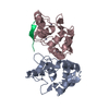
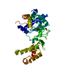
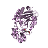
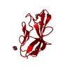
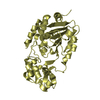
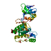
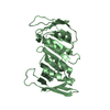
AUTHORS STATE THAT CRYSTAL PACKING ANALYSIS AND ANALYTICAL SIZE EXCLUSION CHROMATOGRAPHY SUPPORT THE ASSIGNMENT OF A DIMER AS A SIGNIFICANT OLIGOMERIC STATE.
-
Components
#1: Protein
putativeglucansynthesisregulatorofSmi1/Knr4family
Mass: 16551.195 Da / Num. of mol.: 2 Source method: isolated from a genetically manipulated source Source: (gene. exp.) Bacteroides fragilis NCTC 9343 (bacteria) Gene: YP_211376.1, BF1740 / Plasmid: SpeedET / Production host: Escherichia Coli (E. coli) / Strain (production host): HK100 / References: UniProt: Q5LEL2
Mass: 18.015 Da / Num. of mol.: 335 / Source method: isolated from a natural source / Formula: H2O
Has protein modification
Y
Sequence details
THE CONSTRUCT WAS EXPRESSED WITH A PURIFICATION TAG MGSDKIHHHHHHENLYFQG. THE TAG WAS REMOVED WITH ...THE CONSTRUCT WAS EXPRESSED WITH A PURIFICATION TAG MGSDKIHHHHHHENLYFQG. THE TAG WAS REMOVED WITH TEV PROTEASE LEAVING ONLY A GLYCINE (0) FOLLOWED BY THE TARGET SEQUENCE.
-
Experimental details
-
Experiment
Experiment
Method: X-RAY DIFFRACTION / Number of used crystals: 1
-
Sample preparation
Crystal
Density Matthews: 2.25 Å3/Da / Density % sol: 45.31 %
Crystal grow
Temperature: 277 K / Method: vapor diffusion, sitting drop / pH: 5.6 Details: 15.0000% Glycerol, 0.1700M NH4OAc, 25.5000% PEG-4000, 0.1M Citrate pH 5.6, NANODROP, VAPOR DIFFUSION, SITTING DROP, temperature 277K
Monochromator: Single crystal Si(111) bent monochromator (horizontal focusing) Protocol: MAD / Monochromatic (M) / Laue (L): M / Scattering type: x-ray
Radiation wavelength
ID
Wavelength (Å)
Relative weight
1
0.91837
1
2
0.97916
1
3
0.97852
1
Reflection
Resolution: 1.45→29.683 Å / Num. obs: 52450 / % possible obs: 98 % / Redundancy: 3.7 % / Biso Wilson estimate: 15.17 Å2 / Rmerge(I) obs: 0.065 / Rsym value: 0.065 / Net I/σ(I): 7.6
Reflection shell
Diffraction-ID: 1
Resolution (Å)
Redundancy (%)
Rmerge(I) obs
Mean I/σ(I) obs
Num. measured all
Num. unique all
Rsym value
% possible all
1.45-1.49
3.3
0.558
1.4
10646
3206
0.558
83
1.49-1.53
3.6
0.483
1.6
13504
3705
0.483
97.5
1.53-1.57
3.7
0.396
1.9
13615
3667
0.396
98.6
1.57-1.62
3.7
0.326
2.3
13236
3553
0.326
98.7
1.62-1.67
3.7
0.259
2.9
12844
3465
0.259
99.1
1.67-1.73
3.7
0.218
3.5
12499
3354
0.218
98.8
1.73-1.8
3.7
0.176
4.2
11993
3231
0.176
99.2
1.8-1.87
3.7
0.149
5
11600
3122
0.149
99.4
1.87-1.96
3.7
0.113
6.5
11277
3031
0.113
99.4
1.96-2.05
3.7
0.094
7.5
10700
2885
0.094
99.4
2.05-2.16
3.7
0.076
9
10165
2738
0.076
99.7
2.16-2.29
3.7
0.066
10.1
9750
2630
0.066
99.6
2.29-2.45
3.7
0.06
11.5
9046
2445
0.06
99.8
2.45-2.65
3.7
0.055
11.8
8568
2322
0.055
99.8
2.65-2.9
3.7
0.05
12.8
7843
2136
0.05
99.9
2.9-3.24
3.7
0.043
14.3
7134
1939
0.043
99.9
3.24-3.74
3.7
0.039
15.2
6302
1721
0.039
99.5
3.74-4.59
3.6
0.035
16.7
5259
1465
0.035
99.7
4.59-6.48
3.5
0.038
15.8
4137
1176
0.038
99.8
6.48-29.68
3.4
0.04
12.9
2215
659
0.04
96.7
-
Phasing
Phasing
Method: MAD
-
Processing
Software
Name
Version
Classification
NB
REFMAC
5.4.0069
refinement
PHENIX
refinement
SHELX
phasing
MolProbity
3beta29
modelbuilding
SCALA
datascaling
PDB_EXTRACT
3.004
dataextraction
MOSFLM
datareduction
SHELXD
phasing
autoSHARP
phasing
Refinement
Method to determine structure: MAD / Resolution: 1.45→29.683 Å / Cor.coef. Fo:Fc: 0.971 / Cor.coef. Fo:Fc free: 0.967 / SU B: 2.001 / SU ML: 0.039 / TLS residual ADP flag: LIKELY RESIDUAL / Cross valid method: THROUGHOUT / σ(F): 0 / ESU R: 0.062 / ESU R Free: 0.061 Stereochemistry target values: MAXIMUM LIKELIHOOD WITH PHASES Details: 1. HYDROGENS HAVE BEEN ADDED IN THE RIDING POSITIONS. 2. ATOM RECORDS CONTAIN RESIDUAL B FACTORS ONLY. 3. A MET-INHIBITION PROTOCOL WAS USED FOR SELENOMETHIONINE INCORPORATION DURING PROTEIN ...Details: 1. HYDROGENS HAVE BEEN ADDED IN THE RIDING POSITIONS. 2. ATOM RECORDS CONTAIN RESIDUAL B FACTORS ONLY. 3. A MET-INHIBITION PROTOCOL WAS USED FOR SELENOMETHIONINE INCORPORATION DURING PROTEIN EXPRESSION. THE OCCUPANCY OF THE SE ATOMS IN THE MSE RESIDUES WAS REDUCED TO 0.75 FOR THE REDUCED SCATTERING POWER DUE TO PARTIAL S-MET INCORPORATION. 4. ACETATE (ACT) IONS, GLYCEROL (GOL) AND PEG4000 FRAGMENT (PEG) MOLECULES FROM CRYSTALLIZATION WERE MODELED.
Rfactor
Num. reflection
% reflection
Selection details
Rfree
0.175
2676
5.1 %
RANDOM
Rwork
0.156
-
-
-
obs
0.157
52409
97.72 %
-
Solvent computation
Ion probe radii: 0.8 Å / Shrinkage radii: 0.8 Å / VDW probe radii: 1.2 Å / Solvent model: MASK
Displacement parameters
Biso mean: 13.475 Å2
Baniso -1
Baniso -2
Baniso -3
1-
0.11 Å2
0 Å2
0 Å2
2-
-
-0.3 Å2
0 Å2
3-
-
-
0.19 Å2
Refinement step
Cycle: LAST / Resolution: 1.45→29.683 Å
Protein
Nucleic acid
Ligand
Solvent
Total
Num. atoms
2138
0
37
335
2510
Refine LS restraints
Refine-ID
Type
Dev ideal
Dev ideal target
Number
X-RAY DIFFRACTION
r_bond_refined_d
0.017
0.022
2474
X-RAY DIFFRACTION
r_bond_other_d
0.001
0.02
1618
X-RAY DIFFRACTION
r_angle_refined_deg
1.677
1.956
3374
X-RAY DIFFRACTION
r_angle_other_deg
1.418
3
3974
X-RAY DIFFRACTION
r_dihedral_angle_1_deg
5.803
5
317
X-RAY DIFFRACTION
r_dihedral_angle_2_deg
32.071
25.478
115
X-RAY DIFFRACTION
r_dihedral_angle_3_deg
14.591
15
412
X-RAY DIFFRACTION
r_dihedral_angle_4_deg
20.154
15
3
X-RAY DIFFRACTION
r_chiral_restr
0.107
0.2
346
X-RAY DIFFRACTION
r_gen_planes_refined
0.01
0.02
2880
X-RAY DIFFRACTION
r_gen_planes_other
0.003
0.02
519
X-RAY DIFFRACTION
r_mcbond_it
1.369
2
1488
X-RAY DIFFRACTION
r_mcbond_other
0.375
2
612
X-RAY DIFFRACTION
r_mcangle_it
2.25
3
2403
X-RAY DIFFRACTION
r_scbond_it
1.847
2
986
X-RAY DIFFRACTION
r_scangle_it
2.718
3
971
LS refinement shell
Resolution: 1.45→1.488 Å / Total num. of bins used: 20
Rfactor
Num. reflection
% reflection
Rfree
0.234
181
-
Rwork
0.238
3020
-
all
-
3201
-
obs
-
-
82.01 %
Refinement TLS params.
Method: refined / Refine-ID: X-RAY DIFFRACTION
ID
L11 (°2)
L12 (°2)
L13 (°2)
L22 (°2)
L23 (°2)
L33 (°2)
S11 (Å °)
S12 (Å °)
S13 (Å °)
S21 (Å °)
S22 (Å °)
S23 (Å °)
S31 (Å °)
S32 (Å °)
S33 (Å °)
T11 (Å2)
T12 (Å2)
T13 (Å2)
T22 (Å2)
T23 (Å2)
T33 (Å2)
Origin x (Å)
Origin y (Å)
Origin z (Å)
1
0.4467
-0.1259
-0.0366
1.2399
0.2917
0.4093
-0.0145
0.0166
0.003
-0.1295
-0.0632
0.1283
-0.0932
-0.0944
0.0777
-0.0115
0.0177
-0.0268
-0.0176
-0.0183
-0.0253
47.0064
42.3661
12.3956
2
0.3904
0.0578
0.0826
0.5432
0.0951
0.3584
0.001
-0.0028
-0.048
0.038
-0.0126
0.0377
0.0088
-0.0407
0.0116
-0.0218
-0.0041
-0.0033
-0.0073
-0.0064
-0.0145
50.073
16.9378
9.1665
Refinement TLS group
ID
Refine-ID
Refine TLS-ID
Auth asym-ID
Label asym-ID
Auth seq-ID
Label seq-ID
1
X-RAY DIFFRACTION
1
A
A
0 - 132
1 - 133
2
X-RAY DIFFRACTION
2
B
B
0 - 132
1 - 133
+
About Yorodumi
-
News
-
Feb 9, 2022. New format data for meta-information of EMDB entries
New format data for meta-information of EMDB entries
Version 3 of the EMDB header file is now the official format.
The previous official version 1.9 will be removed from the archive.
In the structure databanks used in Yorodumi, some data are registered as the other names, "COVID-19 virus" and "2019-nCoV". Here are the details of the virus and the list of structure data.
Jan 31, 2019. EMDB accession codes are about to change! (news from PDBe EMDB page)
EMDB accession codes are about to change! (news from PDBe EMDB page)
The allocation of 4 digits for EMDB accession codes will soon come to an end. Whilst these codes will remain in use, new EMDB accession codes will include an additional digit and will expand incrementally as the available range of codes is exhausted. The current 4-digit format prefixed with “EMD-” (i.e. EMD-XXXX) will advance to a 5-digit format (i.e. EMD-XXXXX), and so on. It is currently estimated that the 4-digit codes will be depleted around Spring 2019, at which point the 5-digit format will come into force.
The EM Navigator/Yorodumi systems omit the EMD- prefix.
Related info.:Q: What is EMD? / ID/Accession-code notation in Yorodumi/EM Navigator
Yorodumi is a browser for structure data from EMDB, PDB, SASBDB, etc.
This page is also the successor to EM Navigator detail page, and also detail information page/front-end page for Omokage search.
The word "yorodu" (or yorozu) is an old Japanese word meaning "ten thousand". "mi" (miru) is to see.
Related info.:EMDB / PDB / SASBDB / Comparison of 3 databanks / Yorodumi Search / Aug 31, 2016. New EM Navigator & Yorodumi / Yorodumi Papers / Jmol/JSmol / Function and homology information / Changes in new EM Navigator and Yorodumi
 Movie
Movie Controller

 Yorodumi
 Open data
 Basic information
 Components
 Keywords
 Function and homology information
 Bacteroides fragilis NCTC 9343 (bacteria)
 X-RAY DIFFRACTION /
  Authors
 Citation
 Structure visualization
 Downloads & links
 Other downloads
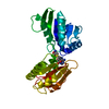


 PDBj
PDBj Assembly

 Mass: 59.044 Da / Num. of mol.: 3 / Source method: obtained synthetically / Formula: C2H3O2
 Mass: 92.094 Da / Num. of mol.: 3 / Source method: obtained synthetically / Formula: C3H8O3
 Mass: 106.120 Da / Num. of mol.: 1 / Source method: obtained synthetically / Formula: C4H10O3
 Mass: 18.015 Da / Num. of mol.: 335 / Source method: isolated from a natural source / Formula: H2O
 Sample preparation
 / Beamline: BL11-1 / Wavelength: 0.91837,0.97916,0.97852
 Processing