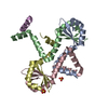
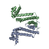
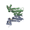
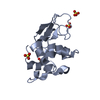
- PDB-3d55: Crystal structure of M. tuberculosis YefM antitoxin -
+
Open data
ID or keywords:
Loading...
-
Basic information
Entry
Database: PDB / ID: 3d55
Title
Crystal structure of M. tuberculosis YefM antitoxin
Components
Uncharacterized protein Rv3357/MT3465
Keywords
TOXIN INHIBITOR / Tetramer / Toxin neutraliser
Function / homology
Function and homology information
sequence-specific DNA binding / DNA-binding transcription factor activity / regulation of DNA-templated transcription / DNA-templated transcription / DNA binding Similarity search - Function
Arc Repressor Mutant - #170 / Single alpha-helices involved in coiled-coils or other helix-helix interfaces - #330 / : / YefM-like domain / Type II toxin-antitoxin system, antitoxin Phd/YefM / Antitoxin Phd_YefM, type II toxin-antitoxin system / YefM-like superfamily / YefM-like fold / Arc Repressor Mutant / Single alpha-helices involved in coiled-coils or other helix-helix interfaces ...Arc Repressor Mutant - #170 / Single alpha-helices involved in coiled-coils or other helix-helix interfaces - #330 / : / YefM-like domain / Type II toxin-antitoxin system, antitoxin Phd/YefM / Antitoxin Phd_YefM, type II toxin-antitoxin system / YefM-like superfamily / YefM-like fold / Arc Repressor Mutant / Single alpha-helices involved in coiled-coils or other helix-helix interfaces / Helix non-globular / Special / Orthogonal Bundle / 3-Layer(aba) Sandwich / Mainly Alpha / Alpha Beta Similarity search - Domain/homology
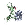
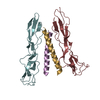
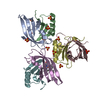
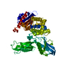
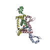
A: Uncharacterized protein Rv3357/MT3465 B: Uncharacterized protein Rv3357/MT3465 C: Uncharacterized protein Rv3357/MT3465 D: Uncharacterized protein Rv3357/MT3465 hetero molecules
Resolution: 2.13→51.3 Å / Cor.coef. Fo:Fc: 0.95 / Cor.coef. Fo:Fc free: 0.939 / SU B: 3.717 / SU ML: 0.096 / Cross valid method: THROUGHOUT / ESU R: 0.046 / ESU R Free: 0.037 / Stereochemistry target values: MAXIMUM LIKELIHOOD Details: HYDROGENS HAVE BEEN ADDED IN THE RIDING POSITIONS; THIS IS A TWINNED DATA. THE TWINNING OPERATOR IS (H,K,L) -> (k, -h, l) AND THE TWINNING FRACTION is 0.404.
Rfactor
Num. reflection
% reflection
Selection details
Rfree
0.21657
969
4.9 %
RANDOM
Rwork
0.17959
-
-
-
obs
0.18151
17054
96.73 %
-
Solvent computation
Ion probe radii: 0.8 Å / Shrinkage radii: 0.8 Å / VDW probe radii: 1.2 Å / Solvent model: MASK
Displacement parameters
Biso mean: 43.252 Å2
Baniso -1
Baniso -2
Baniso -3
1-
6.44 Å2
-11.81 Å2
-0.21 Å2
2-
-
2.93 Å2
0 Å2
3-
-
-
-9.37 Å2
Refinement step
Cycle: LAST / Resolution: 2.13→51.3 Å
Protein
Nucleic acid
Ligand
Solvent
Total
Num. atoms
2549
0
10
56
2615
Refine LS restraints
Refine-ID
Type
Dev ideal
Dev ideal target
Number
X-RAY DIFFRACTION
r_bond_refined_d
0.03
0.021
2575
X-RAY DIFFRACTION
r_angle_refined_deg
2.495
1.957
3482
X-RAY DIFFRACTION
r_dihedral_angle_1_deg
6.627
5
316
X-RAY DIFFRACTION
r_dihedral_angle_2_deg
35.178
23.158
133
X-RAY DIFFRACTION
r_dihedral_angle_3_deg
21.059
15
450
X-RAY DIFFRACTION
r_dihedral_angle_4_deg
16.793
15
33
X-RAY DIFFRACTION
r_chiral_restr
0.233
0.2
388
X-RAY DIFFRACTION
r_gen_planes_refined
0.012
0.021
1970
X-RAY DIFFRACTION
r_mcbond_it
1.687
1.5
1600
X-RAY DIFFRACTION
r_mcangle_it
2.616
2
2570
X-RAY DIFFRACTION
r_scbond_it
4.293
3
975
X-RAY DIFFRACTION
r_scangle_it
6.159
4.5
912
LS refinement shell
Resolution: 2.125→2.18 Å / Total num. of bins used: 20
Rfactor
Num. reflection
% reflection
Rfree
0.218
53
-
Rwork
0.157
882
-
obs
-
1832
63.22 %
+
About Yorodumi
-
News
-
Feb 9, 2022. New format data for meta-information of EMDB entries
New format data for meta-information of EMDB entries
Version 3 of the EMDB header file is now the official format.
The previous official version 1.9 will be removed from the archive.
In the structure databanks used in Yorodumi, some data are registered as the other names, "COVID-19 virus" and "2019-nCoV". Here are the details of the virus and the list of structure data.
Jan 31, 2019. EMDB accession codes are about to change! (news from PDBe EMDB page)
EMDB accession codes are about to change! (news from PDBe EMDB page)
The allocation of 4 digits for EMDB accession codes will soon come to an end. Whilst these codes will remain in use, new EMDB accession codes will include an additional digit and will expand incrementally as the available range of codes is exhausted. The current 4-digit format prefixed with “EMD-” (i.e. EMD-XXXX) will advance to a 5-digit format (i.e. EMD-XXXXX), and so on. It is currently estimated that the 4-digit codes will be depleted around Spring 2019, at which point the 5-digit format will come into force.
The EM Navigator/Yorodumi systems omit the EMD- prefix.
Related info.:Q: What is EMD? / ID/Accession-code notation in Yorodumi/EM Navigator
Yorodumi is a browser for structure data from EMDB, PDB, SASBDB, etc.
This page is also the successor to EM Navigator detail page, and also detail information page/front-end page for Omokage search.
The word "yorodu" (or yorozu) is an old Japanese word meaning "ten thousand". "mi" (miru) is to see.
Related info.:EMDB / PDB / SASBDB / Comparison of 3 databanks / Yorodumi Search / Aug 31, 2016. New EM Navigator & Yorodumi / Yorodumi Papers / Jmol/JSmol / Function and homology information / Changes in new EM Navigator and Yorodumi
 Movie
Movie Controller


 Open data
 Basic information
 Components
 Keywords
 Function and homology information
 Mycobacterium tuberculosis (bacteria)
 X-RAY DIFFRACTION /
  Authors
 Citation
 Structure visualization
 Downloads & links
 Other downloads
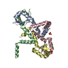
 PDBj
PDBj Assembly
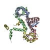


 Mass: 96.063 Da / Num. of mol.: 2 / Source method: obtained synthetically / Formula: SO4
 Mass: 18.015 Da / Num. of mol.: 56 / Source method: isolated from a natural source / Formula: H2O
 Sample preparation
 / Beamline: ID14-2 / Wavelength: 0.933 Å
 Processing