- PDB-3d00: Crystal structure of a tungsten formylmethanofuran dehydrogenase ... -
+
Open data
ID or keywords:
Loading...
-
Basic information
Entry
Database: PDB / ID: 3d00
Title
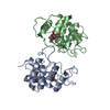
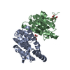
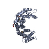
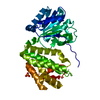
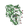
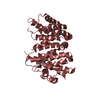
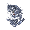
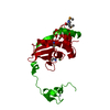
Crystal structure of a tungsten formylmethanofuran dehydrogenase subunit e (fmde)-like protein (syn_00638) from syntrophus aciditrophicus at 1.90 A resolution
Components
Tungsten formylmethanofuran dehydrogenase subunit E
Keywords
METAL BINDING PROTEIN / Fwde/gapdh domain-like fold / structural genomics / Joint Center for Structural Genomics / JCSG / Protein Structure Initiative / PSI-2
AUTHORS STATE THAT THE PROTOMER MAY FORM A DIMER BASED ON CRYSTAL PACKING ANALYSIS. ANALYTICAL SIZE-EXCLUSION CHROMATOGRAPHY SUPPORTS THE ASSIGNMENT OF A DIMER AS THE SIGNIFICANT OLIGOMERIC FORM IN SOLUTION.
-
Components
#1: Protein
TungstenformylmethanofurandehydrogenasesubunitE
Mass: 21697.605 Da / Num. of mol.: 1 Source method: isolated from a genetically manipulated source Source: (gene. exp.) Syntrophus aciditrophicus (bacteria) / Strain: SB / Gene: YP_460196.1, SYNAS_01490, SYN_00638 / Plasmid: SpeedET / Production host: Escherichia coli (E. coli) / Strain (production host): HK100 / References: UniProt: Q2LQ23
Mass: 18.015 Da / Num. of mol.: 42 / Source method: isolated from a natural source / Formula: H2O
Has protein modification
Y
Sequence details
THE CONSTRUCT WAS EXPRESSED WITH A PURIFICATION TAG MGSDKIHHHHHHENLYFQG. THE TAG WAS REMOVED WITH ...THE CONSTRUCT WAS EXPRESSED WITH A PURIFICATION TAG MGSDKIHHHHHHENLYFQG. THE TAG WAS REMOVED WITH TEV PROTEASE LEAVING ONLY A GLYCINE FOLLOWED BY THE TARGET SEQUENCE.
-
Experimental details
-
Experiment
Experiment
Method: X-RAY DIFFRACTION / Number of used crystals: 1
-
Sample preparation
Crystal
Density Matthews: 2.33 Å3/Da / Density % sol: 47.14 %
Crystal grow
Temperature: 277 K / Method: vapor diffusion, sitting drop / pH: 8.5 Details: 0.01M NiCl2, 20.0% PEG MME 2000, 0.1M Tris-HCl pH 8.5, NANODROP, VAPOR DIFFUSION, SITTING DROP, temperature 277K
Monochromator: Single crystal Si(111) bent (horizontal focusing) Protocol: MAD / Monochromatic (M) / Laue (L): M / Scattering type: x-ray
Radiation wavelength
ID
Wavelength (Å)
Relative weight
1
0.91837
1
2
0.97817
1
Reflection
Resolution: 1.9→29.386 Å / Num. obs: 16955 / % possible obs: 99.9 % / Redundancy: 7 % / Biso Wilson estimate: 30.675 Å2 / Rmerge(I) obs: 0.078 / Rsym value: 0.078 / Net I/σ(I): 6.8
Reflection shell
Rmerge(I) obs: 0.011 / Diffraction-ID: 1
Resolution (Å)
Redundancy (%)
Mean I/σ(I) obs
Num. measured all
Num. unique all
Rsym value
% possible all
1.9-1.95
7.1
0.7
8626
1208
1.134
99.9
1.95-2
7.2
0.9
8582
1192
0.855
99.9
2-2.06
7.1
1.2
8253
1157
0.659
100
2.06-2.12
7.1
1.6
8020
1122
0.484
100
2.12-2.19
7.2
1.9
7887
1102
0.41
100
2.19-2.27
7.1
2.3
7549
1068
0.34
100
2.27-2.36
7.2
2.8
7325
1018
0.281
100
2.36-2.45
7.1
3.3
7032
989
0.235
100
2.45-2.56
7.1
4.2
6743
953
0.185
100
2.56-2.69
7.1
5.1
6522
920
0.148
100
2.69-2.83
7
6.1
6142
872
0.122
100
2.83-3
7
7.3
5834
837
0.097
100
3-3.21
7
8.6
5569
796
0.079
100
3.21-3.47
7
10.5
5024
718
0.062
100
3.47-3.8
6.9
13.2
4748
693
0.049
100
3.8-4.25
6.8
13.6
4272
631
0.044
100
4.25-4.91
6.7
14.8
3748
561
0.041
100
4.91-6.01
6.5
16
3173
485
0.041
100
6.01-8.5
6
14.3
2353
394
0.041
100
8.5-29.386
5.2
16.4
1244
239
0.035
95.9
-
Phasing
Phasing
Method: MAD
-
Processing
Software
Name
Version
Classification
NB
REFMAC
5.2.0019
refinement
PHENIX
refinement
SHELX
phasing
MolProbity
3beta29
modelbuilding
SCALA
datascaling
PDB_EXTRACT
3.004
dataextraction
MAR345
CCD
datacollection
MOSFLM
datareduction
SHELXD
phasing
autoSHARP
phasing
Refinement
Method to determine structure: MAD / Resolution: 1.9→29.386 Å / Cor.coef. Fo:Fc: 0.946 / Cor.coef. Fo:Fc free: 0.938 / SU B: 11.433 / SU ML: 0.156 / TLS residual ADP flag: LIKELY RESIDUAL / Cross valid method: THROUGHOUT / σ(F): 0 / ESU R: 0.17 / ESU R Free: 0.156 Stereochemistry target values: MAXIMUM LIKELIHOOD WITH PHASES Details: 1. HYDROGENS HAVE BEEN ADDED IN THE RIDING POSITIONS. 2. ATOM RECORD CONTAINS RESIDUAL B FACTORS ONLY. 3. A MET-INHIBITION PROTOCOL WAS USED FOR SELENOMETHIONINE INCORPORATION DURING PROTEIN ...Details: 1. HYDROGENS HAVE BEEN ADDED IN THE RIDING POSITIONS. 2. ATOM RECORD CONTAINS RESIDUAL B FACTORS ONLY. 3. A MET-INHIBITION PROTOCOL WAS USED FOR SELENOMETHIONINE INCORPORATION DURING PROTEIN EXPRESSION. THE OCCUPANCY OF THE SE ATOMS IN THE MSE RESIDUES WAS REDUCED TO 0.75 FOR THE REDUCED SCATTERING POWER DUE TO PARTIAL S-MET INCORPORATION. 4. ZINC WAS MODELED BASED ON X-RAY FLUORESCENCE SCAN AND ANOMALOUS DIFFERENCE FOURIER MAP CALCULATIONS. 5. CHLORIDE WAS MODELED BASED ON CRYSTALLIZATION CONDITIONS.
Rfactor
Num. reflection
% reflection
Selection details
Rfree
0.268
855
5.1 %
RANDOM
Rwork
0.233
-
-
-
obs
0.235
16902
99.9 %
-
Solvent computation
Ion probe radii: 0.8 Å / Shrinkage radii: 0.8 Å / VDW probe radii: 1.2 Å / Solvent model: MASK
Displacement parameters
Biso mean: 35.2 Å2
Baniso -1
Baniso -2
Baniso -3
1-
1.73 Å2
0 Å2
0 Å2
2-
-
1.73 Å2
0 Å2
3-
-
-
-3.46 Å2
Refinement step
Cycle: LAST / Resolution: 1.9→29.386 Å
Protein
Nucleic acid
Ligand
Solvent
Total
Num. atoms
1388
0
2
42
1432
Refine LS restraints
Refine-ID
Type
Dev ideal
Dev ideal target
Number
X-RAY DIFFRACTION
r_bond_refined_d
0.019
0.022
1445
X-RAY DIFFRACTION
r_bond_other_d
0.003
0.02
961
X-RAY DIFFRACTION
r_angle_refined_deg
1.604
1.987
1972
X-RAY DIFFRACTION
r_angle_other_deg
1.031
3
2356
X-RAY DIFFRACTION
r_dihedral_angle_1_deg
6.4
5
189
X-RAY DIFFRACTION
r_dihedral_angle_2_deg
34.474
23.774
53
X-RAY DIFFRACTION
r_dihedral_angle_3_deg
14.661
15
225
X-RAY DIFFRACTION
r_dihedral_angle_4_deg
21.608
15
6
X-RAY DIFFRACTION
r_chiral_restr
0.088
0.2
224
X-RAY DIFFRACTION
r_gen_planes_refined
0.006
0.02
1615
X-RAY DIFFRACTION
r_gen_planes_other
0.001
0.02
292
X-RAY DIFFRACTION
r_nbd_refined
0.2
0.2
285
X-RAY DIFFRACTION
r_nbd_other
0.196
0.2
900
X-RAY DIFFRACTION
r_nbtor_refined
0.182
0.2
710
X-RAY DIFFRACTION
r_nbtor_other
0.087
0.2
747
X-RAY DIFFRACTION
r_xyhbond_nbd_refined
0.175
0.2
37
X-RAY DIFFRACTION
r_symmetry_vdw_refined
0.114
0.2
15
X-RAY DIFFRACTION
r_symmetry_vdw_other
0.226
0.2
37
X-RAY DIFFRACTION
r_symmetry_hbond_refined
0.178
0.2
15
X-RAY DIFFRACTION
r_mcbond_it
1.214
2
954
X-RAY DIFFRACTION
r_mcbond_other
0.264
2
376
X-RAY DIFFRACTION
r_mcangle_it
1.913
3
1489
X-RAY DIFFRACTION
r_scbond_it
1.086
2
576
X-RAY DIFFRACTION
r_scangle_it
1.576
3
480
LS refinement shell
Resolution: 1.9→1.949 Å / Total num. of bins used: 20
Rfactor
Num. reflection
% reflection
Rfree
0.351
64
-
Rwork
0.347
1136
-
all
-
1200
-
obs
-
-
99.75 %
Refinement TLS params.
Method: refined / Origin x: 16.6441 Å / Origin y: 25.3781 Å / Origin z: 61.974 Å
11
12
13
21
22
23
31
32
33
T
-0.1523 Å2
-0.0519 Å2
0.0003 Å2
-
-0.1421 Å2
0.0085 Å2
-
-
-0.1545 Å2
L
1.5378 °2
-1.5082 °2
2.3567 °2
-
3.3908 °2
-3.3548 °2
-
-
5.9005 °2
S
-0.0174 Å °
0.2052 Å °
0.1697 Å °
0.1243 Å °
-0.1391 Å °
-0.1022 Å °
-0.3315 Å °
0.008 Å °
0.1565 Å °
+
About Yorodumi
-
News
-
Feb 9, 2022. New format data for meta-information of EMDB entries
New format data for meta-information of EMDB entries
Version 3 of the EMDB header file is now the official format.
The previous official version 1.9 will be removed from the archive.
In the structure databanks used in Yorodumi, some data are registered as the other names, "COVID-19 virus" and "2019-nCoV". Here are the details of the virus and the list of structure data.
Jan 31, 2019. EMDB accession codes are about to change! (news from PDBe EMDB page)
EMDB accession codes are about to change! (news from PDBe EMDB page)
The allocation of 4 digits for EMDB accession codes will soon come to an end. Whilst these codes will remain in use, new EMDB accession codes will include an additional digit and will expand incrementally as the available range of codes is exhausted. The current 4-digit format prefixed with “EMD-” (i.e. EMD-XXXX) will advance to a 5-digit format (i.e. EMD-XXXXX), and so on. It is currently estimated that the 4-digit codes will be depleted around Spring 2019, at which point the 5-digit format will come into force.
The EM Navigator/Yorodumi systems omit the EMD- prefix.
Related info.:Q: What is EMD? / ID/Accession-code notation in Yorodumi/EM Navigator
Yorodumi is a browser for structure data from EMDB, PDB, SASBDB, etc.
This page is also the successor to EM Navigator detail page, and also detail information page/front-end page for Omokage search.
The word "yorodu" (or yorozu) is an old Japanese word meaning "ten thousand". "mi" (miru) is to see.
Related info.:EMDB / PDB / SASBDB / Comparison of 3 databanks / Yorodumi Search / Aug 31, 2016. New EM Navigator & Yorodumi / Yorodumi Papers / Jmol/JSmol / Function and homology information / Changes in new EM Navigator and Yorodumi
 Movie
Movie Controller

 Yorodumi
 Open data
 Basic information
 Components
 Keywords
 Function and homology information
 Syntrophus aciditrophicus (bacteria)
 X-RAY DIFFRACTION /
  Authors
 Citation
 Structure visualization
 Downloads & links
 Other downloads

 PDBj
PDBj Assembly
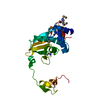

 Mass: 65.409 Da / Num. of mol.: 1 / Source method: obtained synthetically / Formula: Zn
 Mass: 35.453 Da / Num. of mol.: 1 / Source method: obtained synthetically / Formula: Cl
 Mass: 18.015 Da / Num. of mol.: 42 / Source method: isolated from a natural source / Formula: H2O
 Sample preparation
 / Beamline: BL11-1 / Wavelength: 0.91837,0.97817
 Processing