SEQUENCE THE CONSTRUCT WAS EXPRESSED WITH A PURIFICATION TAG: MGSDKIHHHHHHENLYFQG. THE TAG WAS ...SEQUENCE THE CONSTRUCT WAS EXPRESSED WITH A PURIFICATION TAG: MGSDKIHHHHHHENLYFQG. THE TAG WAS REMOVED WITH TEV PROTEASE LEAVING ONLY GLYCINE (0) FOLLOWED BY THE TARGET SEQUENCE.
Mass: 18.015 Da / Num. of mol.: 129 / Source method: isolated from a natural source / Formula: H2O
-
Details
Has protein modification
Y
-
Experimental details
-
Experiment
Experiment
Method: X-RAY DIFFRACTION / Number of used crystals: 2
-
Sample preparation
Crystal
ID
Density Matthews (Å3/Da)
Density % sol (%)
Description
1
2.87
56.79
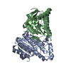
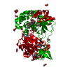
DATA FROM A MONOCLINIC C2 CRYSTAL FORM CONTAINING SE-MET WAS USED FOR THE MAD PHASING EXPERIMENTS AND DETERMINATION OF THE STRUCTURE AT A RESOLUTION OF 2.0 ANGSTROMS. THIS 2.0 ANGSTROM MAD STRUCTURE WAS USED AS A MOLECULAR REPLACEMENT MODEL TO DETERMINE THE STRUCTURE AT AN ENHANCED RESOLUTION OF 1.87 ANGSTROMS IN THE I222 SPACEGROUP.
2
Crystal grow
Temperature (K)
Crystal-ID
Method
pH
Details
277
1
vapor diffusion, sitting drop, nanodrop
6
10.0% PEG-8000, 0.2M Zn(OAc)2, 0.1M MES pH 6.0, VAPOR DIFFUSION,SITTING DROP,NANODROP, temperature 277K
277
2
vapor diffusion, sitting drop, nanodrop
6
0.2M MgNO3, 20.0% PEG-3350, No Buffer pH 5.8, pH 6.0, VAPOR DIFFUSION,SITTING DROP,NANODROP, temperature 277K
-
Data collection
Diffraction
ID
Mean temperature (K)
Crystal-ID
1
100
1
2
100
2
Diffraction source
Source
Site
Beamline
ID
Wavelength (Å)
SYNCHROTRON
SSRL
BL11-1
1
1.000001
SYNCHROTRON
SSRL
BL9-2
2
0.97918, 0.91837, 0.97903
Detector
Type
ID
Detector
Date
Details (eV)
ADSC QUANTUM 315
1
CCD
Mar 19, 2006
Flatmirror (verticalfocusing)
MARMOSAIC 325 mm CCD
2
CCD
Mar 12, 2006
Flatcollimatingmirror, toroidfocusingmirror
Radiation
ID
Protocol
Monochromatic (M) / Laue (L)
Scattering type
Wavelength-ID
Monochromator
1
SINGLEWAVELENGTH
M
x-ray
1
2
MAD
M
x-ray
2
Doublecrystalmonochromator
Radiation wavelength
ID
Wavelength (Å)
Relative weight
1
1.000001
1
2
0.97918
1
3
0.91837
1
4
0.97903
1
Reflection
Resolution: 1.87→30.08 Å / Num. obs: 24246 / % possible obs: 99.5 % / Redundancy: 4.05 % / Biso Wilson estimate: 28.07 Å2 / Rmerge(I) obs: 0.114 / Net I/σ(I): 8.38
Reflection shell
Resolution (Å)
% possible obs (%)
Rmerge(I) obs
Mean I/σ(I) obs
Num. measured obs
Diffraction-ID
% possible all
1.87-1.94
99.5
0.698
1.27
9461
1,2
99.5
1.94-2.01
99.7
0.528
1.67
8346
1,2
2.01-2.11
99.5
0.401
2.23
10016
1,2
2.11-2.22
99.7
0.279
3.12
9164
1,2
2.22-2.36
99.8
0.22
3.92
9319
1,2
2.36-2.54
99.6
0.161
5.18
9107
1,2
2.54-2.79
99.8
0.118
6.94
9212
1,2
2.79-3.19
99.6
0.075
10.02
9283
1,2
3.19-4.02
99.1
0.04
16.86
9349
1,2
4.02-30.1
98.4
0.024
25.78
9441
1,2
-
Phasing
Phasing
Method: MAD, molecular replacement
-
Processing
Software
Name
Version
Classification
NB
REFMAC
5.2.0005
refinement
XSCALE
datascaling
PDB_EXTRACT
1.701
dataextraction
XDS
datareduction
SHELXD
phasing
autoSHARP
phasing
PHASER
phasing
Refinement
Method to determine structure: MAD, MOLECULAR REPLACEMENT / Resolution: 1.87→30.08 Å / Cor.coef. Fo:Fc: 0.953 / Cor.coef. Fo:Fc free: 0.941 / SU B: 5.854 / SU ML: 0.089 / TLS residual ADP flag: LIKELY RESIDUAL / Cross valid method: THROUGHOUT / σ(F): 0 / ESU R: 0.122 / ESU R Free: 0.115 / Stereochemistry target values: MAXIMUM LIKELIHOOD Details: 1). HYDROGENS HAVE BEEN ADDED IN THE RIDING POSITIONS. 2). A MET-INHIBITION PROTOCOL WAS USED FOR SELENOMETHIONINE INCORPORATION DURING PROTEIN EXPRESSION. THE OCCUPANCY OF THE SE ATOMS IN ...Details: 1). HYDROGENS HAVE BEEN ADDED IN THE RIDING POSITIONS. 2). A MET-INHIBITION PROTOCOL WAS USED FOR SELENOMETHIONINE INCORPORATION DURING PROTEIN EXPRESSION. THE OCCUPANCY OF THE SE ATOMS IN THE MSE RESIDUES WAS REDUCED TO 0.75 TO ACCOUNT FOR THE REDUCED SCATTERING POWER DUE TO PARTIAL S-MET INCORPORATION. 3). GLYCEROL, ACETATE, AND ZINC CATIONS ARE MODELED BASED ON THE CRYSTALLIZATION CONDITIONS. 4). X-RAY ANOMALOUS SCATTERING MEASUREMENTS INDICATE ZINC CATIONS ARE COORDINATED TO HIS AND CYS SIDECHAIN ATOMS. 5). ATOM RECORD CONTAINS RESIDUAL B FACTORS 6). ELECTRON DENSITY INDICATED THAT THE SIDECHAIN SULFUR ATOM OF OF CYS 61 NEAR THE PUTATIVE ACTIVE SITE IS IN COVALENT BONDING DISTANCE OF AN UNKNOWN MOLECULE, AND AN UNKNOWN LIGAND (UNL) WAS MODELED AT THIS POSITION.
Rfactor
Num. reflection
% reflection
Selection details
Rfree
0.217
1229
5.1 %
RANDOM
Rwork
0.19
-
-
-
all
0.191
-
-
-
obs
0.19109
24246
99.66 %
-
Solvent computation
Ion probe radii: 0.8 Å / Shrinkage radii: 0.8 Å / VDW probe radii: 1.2 Å / Solvent model: BABINET MODEL WITH MASK
Displacement parameters
Biso mean: 31.123 Å2
Baniso -1
Baniso -2
Baniso -3
1-
0.56 Å2
0 Å2
0 Å2
2-
-
0.9 Å2
0 Å2
3-
-
-
-1.46 Å2
Refinement step
Cycle: LAST / Resolution: 1.87→30.08 Å
Protein
Nucleic acid
Ligand
Solvent
Total
Num. atoms
1582
0
68
129
1779
Refine LS restraints
Refine-ID
Type
Dev ideal
Dev ideal target
Number
X-RAY DIFFRACTION
r_bond_refined_d
0.014
0.022
1710
X-RAY DIFFRACTION
r_bond_other_d
0.003
0.02
1533
X-RAY DIFFRACTION
r_angle_refined_deg
1.644
1.989
2279
X-RAY DIFFRACTION
r_angle_other_deg
1.131
3
3553
X-RAY DIFFRACTION
r_dihedral_angle_1_deg
3.609
5
207
X-RAY DIFFRACTION
r_dihedral_angle_2_deg
29.197
23.117
77
X-RAY DIFFRACTION
r_dihedral_angle_3_deg
10.225
15
272
X-RAY DIFFRACTION
r_dihedral_angle_4_deg
9.874
15
11
X-RAY DIFFRACTION
r_chiral_restr
0.097
0.2
225
X-RAY DIFFRACTION
r_gen_planes_refined
0.005
0.02
1906
X-RAY DIFFRACTION
r_gen_planes_other
0.002
0.02
367
X-RAY DIFFRACTION
r_nbd_refined
0.179
0.3
298
X-RAY DIFFRACTION
r_nbd_other
0.125
0.3
1452
X-RAY DIFFRACTION
r_nbtor_refined
0.177
0.5
804
X-RAY DIFFRACTION
r_nbtor_other
0.081
0.5
886
X-RAY DIFFRACTION
r_xyhbond_nbd_refined
0.153
0.5
165
X-RAY DIFFRACTION
r_metal_ion_refined
0.144
0.5
2
X-RAY DIFFRACTION
r_symmetry_vdw_refined
0.14
0.3
38
X-RAY DIFFRACTION
r_symmetry_vdw_other
0.141
0.3
111
X-RAY DIFFRACTION
r_symmetry_hbond_refined
0.22
0.5
43
X-RAY DIFFRACTION
r_symmetry_metal_ion_refined
0.09
0.5
1
X-RAY DIFFRACTION
r_mcbond_it
1.53
3
1075
X-RAY DIFFRACTION
r_mcbond_other
0.328
3
419
X-RAY DIFFRACTION
r_mcangle_it
2.323
5
1627
X-RAY DIFFRACTION
r_scbond_it
3.861
8
753
X-RAY DIFFRACTION
r_scangle_it
5.003
11
649
LS refinement shell
Resolution: 1.87→1.919 Å / Total num. of bins used: 20
Rfactor
Num. reflection
% reflection
Rfree
0.27
77
-
Rwork
0.279
1671
-
obs
-
1748
99.77 %
Refinement TLS params.
Method: refined / Origin x: 33.842 Å / Origin y: 19.94 Å / Origin z: 4.083 Å
11
12
13
21
22
23
31
32
33
T
-0.1532 Å2
0.0103 Å2
-0.0024 Å2
-
-0.1698 Å2
-0.0107 Å2
-
-
-0.1275 Å2
L
1.5761 °2
-0.5123 °2
-0.5528 °2
-
0.5735 °2
0.1213 °2
-
-
0.7577 °2
S
-0.0546 Å °
-0.0905 Å °
0.1003 Å °
0.0103 Å °
0.0476 Å °
-0.0118 Å °
-0.101 Å °
-0.0208 Å °
0.007 Å °
Refinement TLS group
Selection: ALL
+
About Yorodumi
-
News
-
Feb 9, 2022. New format data for meta-information of EMDB entries
New format data for meta-information of EMDB entries
Version 3 of the EMDB header file is now the official format.
The previous official version 1.9 will be removed from the archive.
In the structure databanks used in Yorodumi, some data are registered as the other names, "COVID-19 virus" and "2019-nCoV". Here are the details of the virus and the list of structure data.
Jan 31, 2019. EMDB accession codes are about to change! (news from PDBe EMDB page)
EMDB accession codes are about to change! (news from PDBe EMDB page)
The allocation of 4 digits for EMDB accession codes will soon come to an end. Whilst these codes will remain in use, new EMDB accession codes will include an additional digit and will expand incrementally as the available range of codes is exhausted. The current 4-digit format prefixed with “EMD-” (i.e. EMD-XXXX) will advance to a 5-digit format (i.e. EMD-XXXXX), and so on. It is currently estimated that the 4-digit codes will be depleted around Spring 2019, at which point the 5-digit format will come into force.
The EM Navigator/Yorodumi systems omit the EMD- prefix.
Related info.:Q: What is EMD? / ID/Accession-code notation in Yorodumi/EM Navigator
Yorodumi is a browser for structure data from EMDB, PDB, SASBDB, etc.
This page is also the successor to EM Navigator detail page, and also detail information page/front-end page for Omokage search.
The word "yorodu" (or yorozu) is an old Japanese word meaning "ten thousand". "mi" (miru) is to see.
Related info.:EMDB / PDB / SASBDB / Comparison of 3 databanks / Yorodumi Search / Aug 31, 2016. New EM Navigator & Yorodumi / Yorodumi Papers / Jmol/JSmol / Function and homology information / Changes in new EM Navigator and Yorodumi
 Movie
Movie Controller

 Yorodumi
 Open data
 Basic information
 Components
 Keywords
 Function and homology information
 Thermoplasma acidophilum (acidophilic)
 X-RAY DIFFRACTION /
  Authors
 Citation
 Structure visualization
 Downloads & links
 Other downloads


 PDBj
PDBj


 Assembly


 Mass: 65.409 Da / Num. of mol.: 5 / Source method: obtained synthetically / Formula: Zn
 Mass: 62.068 Da / Num. of mol.: 6 / Source method: obtained synthetically / Formula: C2H6O2
 Mass: 60.052 Da / Num. of mol.: 8 / Source method: obtained synthetically / Formula: C2H4O2
 Sample preparation
 Processing