- PDB-2glz: Crystal structure of a formylmethanofuran dehydrogenase subunit e... -
+
Open data
ID or keywords:
Loading...
-
Basic information
Entry
Database: PDB / ID: 2glz
Title
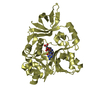
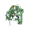
Crystal structure of a formylmethanofuran dehydrogenase subunit e-like protein (dhaf_2992) from desulfitobacterium hafniense dcb-2 at 1.45 A resolution
Components
similar to Formylmethanofuran dehydrogenase subunit E
Keywords
METAL BINDING PROTEIN / Structural genomics / Joint Center for Structural Genomics / JCSG / Protein Structure Initiative / PSI-2
SEQUENCE THE CONSTRUCT WAS EXPRESSED WITH A PURIFICATION TAG: MGSDKIHHHHHHENLYFQG. THE TAG WAS ...SEQUENCE THE CONSTRUCT WAS EXPRESSED WITH A PURIFICATION TAG: MGSDKIHHHHHHENLYFQG. THE TAG WAS REMOVED WITH TEV PROTEASE LEAVING ONLY GLYCINE (0) FOLLOWED BY THE TARGET SEQUENCE.
Resolution: 1.45→29.894 Å / Num. obs: 71200 / % possible obs: 99.9 % / Redundancy: 7.4 % / Biso Wilson estimate: 18.1 Å2 / Rmerge(I) obs: 0.071 / Rsym value: 0.071 / Net I/σ(I): 5.8
Reflection shell
Diffraction-ID: 1
Resolution (Å)
Redundancy (%)
Rmerge(I) obs
Mean I/σ(I) obs
Num. measured all
Num. unique obs
Rsym value
% possible all
1.45-1.49
4.6
0.729
1
24112
5186
0.729
99.9
1.49-1.53
7.4
0.63
1
37182
5034
0.63
99.9
1.53-1.57
7.6
0.493
1.5
37810
4965
0.493
100
1.57-1.62
7.6
0.374
2
36437
4772
0.374
100
1.62-1.67
7.6
0.298
2.5
35494
4652
0.298
100
1.67-1.73
7.6
0.257
2.9
34680
4545
0.257
100
1.73-1.8
7.7
0.201
3.7
33160
4329
0.201
100
1.8-1.87
7.7
0.161
4.5
32179
4192
0.161
100
1.87-1.96
7.6
0.145
4
30513
4026
0.145
100
1.96-2.05
7.7
0.111
6
29699
3877
0.111
100
2.05-2.16
7.6
0.102
6
27621
3656
0.102
100
2.16-2.29
7.5
0.09
6.5
26497
3516
0.09
100
2.29-2.45
7.7
0.074
8.1
25265
3291
0.074
100
2.45-2.65
7.7
0.066
8.8
23432
3059
0.066
100
2.65-2.9
7.6
0.058
10.4
21842
2856
0.058
100
2.9-3.24
7.6
0.052
11.5
19586
2574
0.052
100
3.24-3.74
7.5
0.05
11.6
17262
2287
0.05
100
3.74-4.59
7.5
0.047
12.9
14671
1968
0.047
99.8
4.59-6.48
7.3
0.045
12.9
11220
1545
0.045
99.8
6.48-29.89
6.5
0.045
12.2
5688
870
0.045
94.8
-
Phasing
Phasing
Method: MAD
-
Processing
Software
Name
Version
Classification
NB
REFMAC
5.2.0005
refinement
SCALA
datascaling
PDB_EXTRACT
1.701
dataextraction
MOSFLM
datareduction
CCP4
(SCALA)
datascaling
SOLVE
phasing
Refinement
Method to determine structure: MAD / Resolution: 1.45→27.2 Å / Cor.coef. Fo:Fc: 0.971 / Cor.coef. Fo:Fc free: 0.96 / SU B: 2.688 / SU ML: 0.05 / TLS residual ADP flag: LIKELY RESIDUAL / Cross valid method: THROUGHOUT / σ(F): 0 / ESU R: 0.058 / ESU R Free: 0.061 Stereochemistry target values: MAXIMUM LIKELIHOOD WITH PHASES Details: 1). HYDROGENS HAVE BEEN ADDED IN THE RIDING POSITIONS. 2). A MET-INHIBITION PROTOCOL WAS USED FOR SELENOMETHIONINE INCORPORATION DURING PROTEIN EXPRESSION. THE OCCUPANCY OF THE SE ATOMS IN ...Details: 1). HYDROGENS HAVE BEEN ADDED IN THE RIDING POSITIONS. 2). A MET-INHIBITION PROTOCOL WAS USED FOR SELENOMETHIONINE INCORPORATION DURING PROTEIN EXPRESSION. THE OCCUPANCY OF THE SE ATOMS IN THE MSE RESIDUES WAS REDUCED TO 0.75 TO ACCOUNT FOR THE REDUCED SCATTERING POWER DUE TO PARTIAL S-MET INCORPORATION. 3). X-RAY ANOMALOUS SCATTERING MEASUREMENTS INDICATE EITHER A ZINC OR A NICKEL CATION ION IS COORDINATED IN AN TETRAHEDERAL COMPLEX TO THE SIDECHAINS OF HIS-15, HIS-17, CYS-19, AND CYS-55. THE RELATIVE OCCUPANCIES OF THE THE METAL IONS WERE ESTIMATED FROM THE RATIO OF THEIR ANOMALOUS DIFFERENCE MAP PEAK HEIGHTS. THE TOTAL OCCUPANCY OF THE ZN AND NI CATIONS WAS REDUCED TO 0.75 TO ACCOUNT FOR THE REDUCED SCATTERING OBSERVED AT THIS SITE. 4). SEVERAL MOLECULES OF THE CRYOPROTECTANT, ETHYLENE GLYCOL, WERE MODELED INTO THE STRUCTURE. 5). UNKNOWN DIFFERENCE DENSITY, BETWEEN THE SIDECHAINS OF ASN 64 AND PHE 87 ON BOTH SUBUNITS, IN THE ASYMMETRIC UNIT HAS NOT BEEN MODELED. 6). ATOM RECORD CONTAINS RESIDUAL B FACTORS ONLY.
Rfactor
Num. reflection
% reflection
Selection details
Rfree
0.198
3593
5.1 %
RANDOM
Rwork
0.171
-
-
-
obs
0.173
71126
99.86 %
-
Solvent computation
Ion probe radii: 0.8 Å / Shrinkage radii: 0.8 Å / VDW probe radii: 1.2 Å / Solvent model: BABINET MODEL WITH MASK
Displacement parameters
Biso mean: 27.137 Å2
Baniso -1
Baniso -2
Baniso -3
1-
2.49 Å2
0 Å2
0 Å2
2-
-
-1.5 Å2
0 Å2
3-
-
-
-0.99 Å2
Refinement step
Cycle: LAST / Resolution: 1.45→27.2 Å
Protein
Nucleic acid
Ligand
Solvent
Total
Num. atoms
2335
0
76
428
2839
Refine LS restraints
Refine-ID
Type
Dev ideal
Dev ideal target
Number
X-RAY DIFFRACTION
r_bond_refined_d
0.017
0.022
2570
X-RAY DIFFRACTION
r_bond_other_d
0.002
0.02
2381
X-RAY DIFFRACTION
r_angle_refined_deg
1.546
1.966
3478
X-RAY DIFFRACTION
r_angle_other_deg
0.807
3
5541
X-RAY DIFFRACTION
r_dihedral_angle_1_deg
5.829
5
329
X-RAY DIFFRACTION
r_dihedral_angle_2_deg
38.092
23.565
115
X-RAY DIFFRACTION
r_dihedral_angle_3_deg
12.204
15
436
X-RAY DIFFRACTION
r_dihedral_angle_4_deg
14.776
15
17
X-RAY DIFFRACTION
r_chiral_restr
0.088
0.2
388
X-RAY DIFFRACTION
r_gen_planes_refined
0.007
0.02
2834
X-RAY DIFFRACTION
r_gen_planes_other
0.002
0.02
509
X-RAY DIFFRACTION
r_nbd_refined
0.21
0.2
502
X-RAY DIFFRACTION
r_nbd_other
0.176
0.2
2431
X-RAY DIFFRACTION
r_nbtor_refined
0.182
0.2
1216
X-RAY DIFFRACTION
r_nbtor_other
0.086
0.2
1477
X-RAY DIFFRACTION
r_xyhbond_nbd_refined
0.158
0.2
305
X-RAY DIFFRACTION
r_metal_ion_refined
0.126
0.2
2
X-RAY DIFFRACTION
r_symmetry_vdw_refined
0.072
0.2
5
X-RAY DIFFRACTION
r_symmetry_vdw_other
0.177
0.2
49
X-RAY DIFFRACTION
r_symmetry_hbond_refined
0.126
0.2
28
X-RAY DIFFRACTION
r_mcbond_it
1.864
3
1590
X-RAY DIFFRACTION
r_mcbond_other
0.503
3
618
X-RAY DIFFRACTION
r_mcangle_it
2.66
5
2526
X-RAY DIFFRACTION
r_scbond_it
4.323
8
1084
X-RAY DIFFRACTION
r_scangle_it
5.661
11
940
LS refinement shell
Resolution: 1.45→1.488 Å / Total num. of bins used: 20
Rfactor
Num. reflection
% reflection
Rfree
0.302
260
-
Rwork
0.27
4921
-
obs
-
5181
99.75 %
Refinement TLS params.
Method: refined / Refine-ID: X-RAY DIFFRACTION
ID
L11 (°2)
L12 (°2)
L13 (°2)
L22 (°2)
L23 (°2)
L33 (°2)
S11 (Å °)
S12 (Å °)
S13 (Å °)
S21 (Å °)
S22 (Å °)
S23 (Å °)
S31 (Å °)
S32 (Å °)
S33 (Å °)
T11 (Å2)
T12 (Å2)
T13 (Å2)
T22 (Å2)
T23 (Å2)
T33 (Å2)
Origin x (Å)
Origin y (Å)
Origin z (Å)
1
1.0424
-0.194
-0.4747
1.2012
-0.1441
1.5542
-0.0072
-0.0403
0.0882
-0.0099
0.0242
-0.025
-0.1092
0.0394
-0.017
-0.2175
-0.005
0.0019
-0.1534
0.0055
-0.1194
32.878
11.877
20.712
2
0.8938
0.2253
0.2856
1.631
0.0699
1.6951
-0.0429
0.0703
-0.0403
-0.0393
0.0677
-0.0395
0.0651
0.0468
-0.0248
-0.2293
-0.0097
-0.0026
-0.1484
0.004
-0.1306
34.649
-12.239
28.211
Refinement TLS group
ID
Refine-ID
Refine TLS-ID
Auth asym-ID
Auth seq-ID
1
X-RAY DIFFRACTION
1
A
3 - 151
2
X-RAY DIFFRACTION
2
B
4 - 151
+
About Yorodumi
-
News
-
Feb 9, 2022. New format data for meta-information of EMDB entries
New format data for meta-information of EMDB entries
Version 3 of the EMDB header file is now the official format.
The previous official version 1.9 will be removed from the archive.
In the structure databanks used in Yorodumi, some data are registered as the other names, "COVID-19 virus" and "2019-nCoV". Here are the details of the virus and the list of structure data.
Jan 31, 2019. EMDB accession codes are about to change! (news from PDBe EMDB page)
EMDB accession codes are about to change! (news from PDBe EMDB page)
The allocation of 4 digits for EMDB accession codes will soon come to an end. Whilst these codes will remain in use, new EMDB accession codes will include an additional digit and will expand incrementally as the available range of codes is exhausted. The current 4-digit format prefixed with “EMD-” (i.e. EMD-XXXX) will advance to a 5-digit format (i.e. EMD-XXXXX), and so on. It is currently estimated that the 4-digit codes will be depleted around Spring 2019, at which point the 5-digit format will come into force.
The EM Navigator/Yorodumi systems omit the EMD- prefix.
Related info.:Q: What is EMD? / ID/Accession-code notation in Yorodumi/EM Navigator
Yorodumi is a browser for structure data from EMDB, PDB, SASBDB, etc.
This page is also the successor to EM Navigator detail page, and also detail information page/front-end page for Omokage search.
The word "yorodu" (or yorozu) is an old Japanese word meaning "ten thousand". "mi" (miru) is to see.
Related info.:EMDB / PDB / SASBDB / Comparison of 3 databanks / Yorodumi Search / Aug 31, 2016. New EM Navigator & Yorodumi / Yorodumi Papers / Jmol/JSmol / Function and homology information / Changes in new EM Navigator and Yorodumi
 Movie
Movie Controller

 Yorodumi
 Open data
 Basic information
 Components
 Keywords
 Function and homology information
 Desulfitobacterium hafniense (bacteria)
 X-RAY DIFFRACTION /
  Authors
 Citation
 Structure visualization
 Downloads & links
 Other downloads


 PDBj
PDBj Assembly
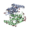

 Mass: 65.409 Da / Num. of mol.: 2 / Source method: obtained synthetically / Formula: Zn
 Mass: 58.693 Da / Num. of mol.: 2 / Source method: obtained synthetically / Formula: Ni
 Mass: 62.068 Da / Num. of mol.: 18 / Source method: obtained synthetically / Formula: C2H6O2
 Mass: 18.015 Da / Num. of mol.: 428 / Source method: isolated from a natural source / Formula: H2O
 Sample preparation
 / Beamline: BL9-2 / Wavelength: 0.91837, 0.97905
 Processing