 Movie
Movie Controller

[English] 日本語
 Yorodumi
- PDB-3cwy: Structure of CagD from H. pylori pathogenicity island crystallize... -
+ Open data

- Basic information
| Entry | Database: PDB / ID: 3cwy | ||||||
|---|---|---|---|---|---|---|---|
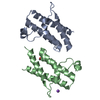
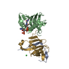
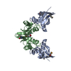
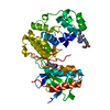
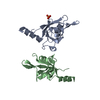
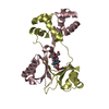
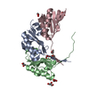
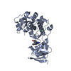
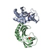
| Title | Structure of CagD from H. pylori pathogenicity island crystallized in the presence of Cu(II) ions | ||||||
 Components Components | protein CagD | ||||||
 Keywords Keywords | UNKNOWN FUNCTION / CagD / cag-pathogenicity island / Type IV secretion system / T4SS | ||||||
| Function / homology |  Function and homology information Function and homology information | ||||||
| Biological species |   Helicobacter pylori (bacteria) Helicobacter pylori (bacteria) | ||||||
| Method |  X-RAY DIFFRACTION / SYNCHROTRON / MOLECULAR REPLACEMENT / Resolution: 2.75 Å X-RAY DIFFRACTION / SYNCHROTRON / MOLECULAR REPLACEMENT / Resolution: 2.75 Å | ||||||
 Authors Authors | Cendron, L. / Zanotti, G. / Angelini, A. / Barison, N. / Couturier, M. / Stein, M. | ||||||
 Citation Citation | Journal: J.Mol.Biol. / Year: 2009 Title: The Helicobacter pylori CagD (HP0545, Cag24) protein is essential for CagA translocation and maximal induction of interleukin-8 secretion. Authors: Cendron, L. / Couturier, M. / Angelini, A. / Barison, N. / Stein, M. / Zanotti, G. #1: Journal: J.Mol.Biol. / Year: 2004Title: Crystal structure of CagZ, a protein from the Helicobacter pylori pathogenicity island that encodes for a type IV secretion system Authors: Cendron, L. / Seydel, A. / Angelini, A. / Battistutta, R. / Zanotti, G. #2: Journal: Proteins: Struct.,Funct.,Genet. / Year: 2007Title: The crystal structure of CagS from the Helicobacter pylori pathogenicity island Authors: Cendron, L. / Tasca, E. / Seraglio, T. / Seydel, A. / Angelini, A. / Battistutta, R. / Montecucco, C. / Zanotti, G. | ||||||
| History |
|
- Structure visualization
| Structure viewer | Molecule: MolmilJmol/JSmol |
|---|
- Downloads & links
-Download
| PDBx/mmCIF format | 3cwy.cif.gz | 41.2 KB | Display | PDBx/mmCIF format |
|---|---|---|---|---|
| PDB format | pdb3cwy.ent.gz | 28.4 KB | Display | PDB format |
| PDBx/mmJSON format | 3cwy.json.gz | Tree view | PDBx/mmJSON format | |
| Others |  Other downloads Other downloads |
-Validation report
| Arichive directory | https://data.pdbj.org/pub/pdb/validation_reports/cw/3cwyftp://data.pdbj.org/pub/pdb/validation_reports/cw/3cwy | HTTPS FTP |
|---|
-Related structure data
| Related structure data | 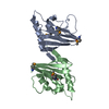 3cwxSC S: Starting model for refinement C: citing same article ( |
|---|---|
| Similar structure data |

-Links
 PDBj
PDBj
- Assembly
| Deposited unit | 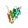
| ||||||||
|---|---|---|---|---|---|---|---|---|---|
| 1 | 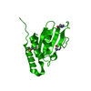
| ||||||||
| Unit cell |
|
-Components
| #1: Protein | Mass: 20777.463 Da / Num. of mol.: 1 / Fragment: CagD / Mutation: V79M, V140M Source method: isolated from a genetically manipulated source Source: (gene. exp.) Helicobacter pylori (bacteria) / Strain: CCUG 17874 / Gene: cagD / Plasmid: pET28b-cagD / Production host: |
|---|---|
| #2: Chemical | ChemComp-CU /   Mass: 63.546 Da / Num. of mol.: 1 / Source method: obtained synthetically / Formula: Cu Mass: 63.546 Da / Num. of mol.: 1 / Source method: obtained synthetically / Formula: Cu |
| #3: Water | ChemComp-HOH /  Mass: 18.015 Da / Num. of mol.: 11 / Source method: isolated from a natural source / Formula: H2O Mass: 18.015 Da / Num. of mol.: 11 / Source method: isolated from a natural source / Formula: H2O |
| Has protein modification | Y |
-Experimental details
-Experiment
| Experiment | Method: X-RAY DIFFRACTION / Number of used crystals: 1 |
|---|
- Sample preparation
| Crystal | Density Matthews: 2.29 Å3/Da / Density % sol: 46.38 % |
|---|---|
| Crystal grow | Temperature: 298 K / Method: vapor diffusion / pH: 7 Details: 0.2 M MgCl2, 0.1 M Hepes pH 7, 20% PEG 6000, CuCl2 0.02M , VAPOR DIFFUSION, temperature 298K |
-Data collection
| Diffraction | Mean temperature: 100 K |
|---|---|
| Diffraction source | Source: SYNCHROTRON / Site: ESRF  / Beamline: BM14 / Wavelength: 0.97621 Å / Beamline: BM14 / Wavelength: 0.97621 Å |
| Detector | Type: MAR CCD 165 mm / Detector: CCD / Date: Aug 31, 2007 |
| Radiation | Protocol: SINGLE WAVELENGTH / Monochromatic (M) / Laue (L): M / Scattering type: x-ray |
| Radiation wavelength | Wavelength: 0.97621 Å / Relative weight: 1 |
| Reflection | Resolution: 2.75→78.09 Å / Num. all: 5757 / Num. obs: 5757 / % possible obs: 99.9 % / Observed criterion σ(F): 0 / Observed criterion σ(I): 0 / Redundancy: 10.3 % / Biso Wilson estimate: 21.5 Å2 / Rmerge(I) obs: 0.079 / Net I/σ(I): 7.5 |
| Reflection shell | Resolution: 2.75→2.85 Å / Redundancy: 10.7 % / Rmerge(I) obs: 0.459 / Mean I/σ(I) obs: 1.7 / Num. unique all: 796 / % possible all: 99 |
- Processing
| Software |
| ||||||||||||||||||||||||||||||||||||
|---|---|---|---|---|---|---|---|---|---|---|---|---|---|---|---|---|---|---|---|---|---|---|---|---|---|---|---|---|---|---|---|---|---|---|---|---|---|
| Refinement | Method to determine structure: MOLECULAR REPLACEMENT Starting model: 3CWX Resolution: 2.75→56.34 Å / Rfactor Rfree error: 0.019 / Data cutoff high absF: 1913839.91 / Data cutoff low absF: 0 / Isotropic thermal model: RESTRAINED / Cross valid method: THROUGHOUT / σ(F): 0 / σ(I): 0
| ||||||||||||||||||||||||||||||||||||
| Solvent computation | Ion probe radii: 0.8 Å / Shrinkage radii: 0.8 Å / VDW probe radii: 1.2 Å / Solvent model: FLAT MODEL / Bsol: 102.977 Å2 / ksol: 0.708042 e/Å3 | ||||||||||||||||||||||||||||||||||||
| Displacement parameters | Biso mean: 56.67 Å2
| ||||||||||||||||||||||||||||||||||||
| Refine analyze |
| ||||||||||||||||||||||||||||||||||||
| Refinement step | Cycle: LAST / Resolution: 2.75→56.34 Å
| ||||||||||||||||||||||||||||||||||||
| Refine LS restraints |
| ||||||||||||||||||||||||||||||||||||
| LS refinement shell | Resolution: 2.75→2.82 Å / Rfactor Rfree error: 0.046 / Total num. of bins used: 20
| ||||||||||||||||||||||||||||||||||||
| Xplor file |
|
