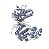
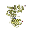
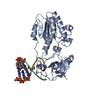
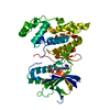
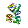
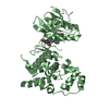
- PDB-3ghd: Crystal structure of a cystathionine beta-synthase domain protein... -
+
Open data
ID or keywords:
Loading...
-
Basic information
Entry
Database: PDB / ID: 3ghd
Title
Crystal structure of a cystathionine beta-synthase domain protein fused to a Zn-ribbon-like domain
Components
a cystathionine beta-synthase domain protein fused to a Zn-ribbon-like domain
Keywords
Nucleotide binding protein / Metal binding protein / PF1953 / APC40009 / cystathionine beta-synthase domain protein / Structural Genomics / PSI-2 / Protein Structure Initiative / Midwest Center for Structural Genomics / MCSG / METAL BINDIN
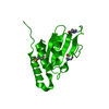
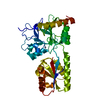
Archaeal CBS protein, (ACP)-type metal binding domain / Archaeal CBS proteins (ACP)-type metal binding (MB) domain profile. / CBS domain Like - #20 / CBS domain Like / : / Domain in cystathionine beta-synthase and other proteins. / CBS domain superfamily / CBS domain / CBS domain / CBS domain profile. ...Archaeal CBS protein, (ACP)-type metal binding domain / Archaeal CBS proteins (ACP)-type metal binding (MB) domain profile. / CBS domain Like - #20 / CBS domain Like / : / Domain in cystathionine beta-synthase and other proteins. / CBS domain superfamily / CBS domain / CBS domain / CBS domain profile. / Alpha-Beta Complex / Alpha Beta Similarity search - Domain/homology
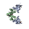
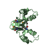
A: a cystathionine beta-synthase domain protein fused to a Zn-ribbon-like domain B: a cystathionine beta-synthase domain protein fused to a Zn-ribbon-like domain
A: a cystathionine beta-synthase domain protein fused to a Zn-ribbon-like domain B: a cystathionine beta-synthase domain protein fused to a Zn-ribbon-like domain
A: a cystathionine beta-synthase domain protein fused to a Zn-ribbon-like domain B: a cystathionine beta-synthase domain protein fused to a Zn-ribbon-like domain
Mass: 18.015 Da / Num. of mol.: 112 / Source method: isolated from a natural source / Formula: H2O
Has protein modification
Y
Sequence details
AUTHORS STATE THAT THE PROTEIN WAS CRYSTALLIZED BY IN SITU PROTEOLYSIS METHOD AND THE EXACT ...AUTHORS STATE THAT THE PROTEIN WAS CRYSTALLIZED BY IN SITU PROTEOLYSIS METHOD AND THE EXACT SEQUENCE WENT INTO THE CRYSTAL WAS NOT DETERMINED. THE PROTEIN WAS MIXED WITH 100:1(W/W) WITH CHYMOTRYPSIN IMMEDIATELY PRIOR TO CRYSTALLIZATION TRIAL. THE FULL SEQUENCE BEFORE PROTEOLYSIS WAS MGSSHHHHHHSSGRENLYFQGMAQKILVEQVVKRKAIVVQPKDTVDRVAKILSRNKAG SAVVMEGDEILGVVTERDILDKVVAKGKNPKEVKVEEIMTKNPVKIEYDYDIEDVIEL MTEKGVRRVLVTKFGKPIGFVTAADILAALASHNHEEEEEEREEESEVYGICEVCGQY GALYKVYHEGRELWVCETCKDLIEGR
-
Experimental details
-
Experiment
Experiment
Method: X-RAY DIFFRACTION / Number of used crystals: 1
-
Sample preparation
Crystal
Density Matthews: 2.25 Å3/Da / Density % sol: 45.44 %
Crystal grow
Temperature: 297 K / Method: vapor diffusion, hanging drop Details: 0.2M Ammonium Chloride,20%PEG3350, VAPOR DIFFUSION, HANGING DROP, temperature 297K
In the structure databanks used in Yorodumi, some data are registered as the other names, "COVID-19 virus" and "2019-nCoV". Here are the details of the virus and the list of structure data.
Jan 31, 2019. EMDB accession codes are about to change! (news from PDBe EMDB page)
EMDB accession codes are about to change! (news from PDBe EMDB page)
The allocation of 4 digits for EMDB accession codes will soon come to an end. Whilst these codes will remain in use, new EMDB accession codes will include an additional digit and will expand incrementally as the available range of codes is exhausted. The current 4-digit format prefixed with “EMD-” (i.e. EMD-XXXX) will advance to a 5-digit format (i.e. EMD-XXXXX), and so on. It is currently estimated that the 4-digit codes will be depleted around Spring 2019, at which point the 5-digit format will come into force.
The EM Navigator/Yorodumi systems omit the EMD- prefix.
Related info.:Q: What is EMD? / ID/Accession-code notation in Yorodumi/EM Navigator
Yorodumi is a browser for structure data from EMDB, PDB, SASBDB, etc.
This page is also the successor to EM Navigator detail page, and also detail information page/front-end page for Omokage search.
The word "yorodu" (or yorozu) is an old Japanese word meaning "ten thousand". "mi" (miru) is to see.
Related info.:EMDB / PDB / SASBDB / Comparison of 3 databanks / Yorodumi Search / Aug 31, 2016. New EM Navigator & Yorodumi / Yorodumi Papers / Jmol/JSmol / Function and homology information / Changes in new EM Navigator and Yorodumi
 Movie
Movie Controller

 Yorodumi
 Open data
 Basic information
 Components
 Keywords
 Function and homology information
 Pyrococcus furiosus (archaea)
 X-RAY DIFFRACTION /
  Authors
 Citation
 Structure visualization
 Downloads & links
 Other downloads

 PDBj
PDBj
 Assembly
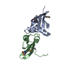

 Mass: 18.015 Da / Num. of mol.: 112 / Source method: isolated from a natural source / Formula: H2O
 Sample preparation
 / Beamline: 23-ID-B / Wavelength: 0.97941 Å
 Processing