 Movie
Movie Controller

[English] 日本語
 Yorodumi
- PDB-3cwx: Crystal structure of cagd from helicobacter pylori pathogenicity ... -
+ Open data

- Basic information
| Entry | Database: PDB / ID: 3cwx | ||||||
|---|---|---|---|---|---|---|---|
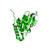
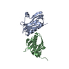
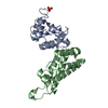
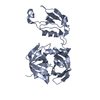
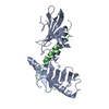
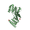
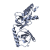
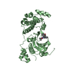
| Title | Crystal structure of cagd from helicobacter pylori pathogenicity island | ||||||
 Components Components | protein CagD | ||||||
 Keywords Keywords | UNKNOWN FUNCTION / CagD / cag-pathogenicity island / Type IV secretion system / T4SS | ||||||
| Function / homology | Pathogenicity island component CagD / Pathogenicity island component CagD / Pathogenicity island component CagD superfamily / Pathogenicity island component CagD / Inhibitor of vertebrate lysozyme, Ivy / 3-Layer(aba) Sandwich / metal ion binding / Alpha Beta / CagD Function and homology information Function and homology information | ||||||
| Biological species |   Helicobacter pylori (bacteria) Helicobacter pylori (bacteria) | ||||||
| Method |  X-RAY DIFFRACTION / SYNCHROTRON / MAD / Resolution: 2.3 Å X-RAY DIFFRACTION / SYNCHROTRON / MAD / Resolution: 2.3 Å | ||||||
 Authors Authors | Cendron, L. / Zanotti, G. / Angelini, A. / Barison, N. / Couturier, M. / Stein, M. | ||||||
 Citation Citation | Journal: J.Mol.Biol. / Year: 2009 Title: The Helicobacter pylori CagD (HP0545, Cag24) protein is essential for CagA translocation and maximal induction of interleukin-8 secretion. Authors: Cendron, L. / Couturier, M. / Angelini, A. / Barison, N. / Stein, M. / Zanotti, G. #1: Journal: J.Mol.Biol. / Year: 2004Title: Crystal structure of CagZ, a protein from the Helicobacter pylori pathogenicity island that encodes for a type IV secretion system Authors: Cendron, L. / Seydel, A. / Angelini, A. / Battistutta, R. / Zanotti, G. #2: Journal: Proteins: Struct.,Funct.,Genet. / Year: 2007Title: The crystal structure of CagS from the Helicobacter pylori pathogenicity island Authors: Cendron, L. / Tasca, E. / Seraglio, T. / Seydel, A. / Angelini, A. / Battistutta, R. / Montecucco, C. / Zanotti, G. | ||||||
| History |
|
- Structure visualization
| Structure viewer | Molecule: MolmilJmol/JSmol |
|---|
- Downloads & links
-Download
| PDBx/mmCIF format | 3cwx.cif.gz | 94.3 KB | Display | PDBx/mmCIF format |
|---|---|---|---|---|
| PDB format | pdb3cwx.ent.gz | 72.3 KB | Display | PDB format |
| PDBx/mmJSON format | 3cwx.json.gz | Tree view | PDBx/mmJSON format | |
| Others |  Other downloads Other downloads |
-Validation report
| Arichive directory | https://data.pdbj.org/pub/pdb/validation_reports/cw/3cwxftp://data.pdbj.org/pub/pdb/validation_reports/cw/3cwx | HTTPS FTP |
|---|
-Related structure data


-Links
 PDBj
PDBj
- Assembly
| Deposited unit | 
| ||||||||
|---|---|---|---|---|---|---|---|---|---|
| 1 | 
| ||||||||
| 2 | 
| ||||||||
| Unit cell |
|
-Components
| #1: Protein | Mass: 20777.463 Da / Num. of mol.: 3 / Fragment: CagD / Mutation: V79M, V140M Source method: isolated from a genetically manipulated source Source: (gene. exp.) Helicobacter pylori (bacteria) / Strain: CCUG 17874 / Gene: cagD / Plasmid: pET28b-cagD / Production host: #2: Water | ChemComp-HOH / |  Mass: 18.015 Da / Num. of mol.: 158 / Source method: isolated from a natural source / Formula: H2O Mass: 18.015 Da / Num. of mol.: 158 / Source method: isolated from a natural source / Formula: H2OHas protein modification | Y | |
|---|
-Experimental details
-Experiment
| Experiment | Method: X-RAY DIFFRACTION / Number of used crystals: 3 |
|---|
- Sample preparation
| Crystal | Density Matthews: 2.26 Å3/Da / Density % sol: 45.6 % |
|---|---|
| Crystal grow | Temperature: 298 K / Method: vapor diffusion / pH: 7 Details: 0.2 M MgGl2, 0.1M Hepes, 20% PEG 6000, pH 7, VAPOR DIFFUSION, temperature 298K |
-Data collection
| Diffraction | Mean temperature: 100 K | ||||||||||||
|---|---|---|---|---|---|---|---|---|---|---|---|---|---|
| Diffraction source | Source: SYNCHROTRON / Site: ESRF  / Beamline: ID14-4 / Wavelength: 0.97942, 0.97420, 0.97814 / Beamline: ID14-4 / Wavelength: 0.97942, 0.97420, 0.97814 | ||||||||||||
| Detector | Type: ADSC QUANTUM 315 / Detector: CCD / Date: Dec 7, 2006 | ||||||||||||
| Radiation | Protocol: MAD / Monochromatic (M) / Laue (L): M / Scattering type: x-ray | ||||||||||||
| Radiation wavelength |
| ||||||||||||
| Reflection | Resolution: 2.2→62.26 Å / Num. all: 28577 / Num. obs: 28577 / % possible obs: 99.2 % / Observed criterion σ(F): 0 / Observed criterion σ(I): 0 / Redundancy: 7.3 % / Rmerge(I) obs: 0.088 / Net I/σ(I): 16.6 | ||||||||||||
| Reflection shell | Resolution: 2.2→2.32 Å / Redundancy: 7.4 % / Rmerge(I) obs: 0.411 / Mean I/σ(I) obs: 4.1 / Num. unique all: 4107 / % possible all: 99.9 |
- Processing
| Software |
| |||||||||||||||||||||||||||||||||
|---|---|---|---|---|---|---|---|---|---|---|---|---|---|---|---|---|---|---|---|---|---|---|---|---|---|---|---|---|---|---|---|---|---|---|
| Refinement | Method to determine structure: MAD / Resolution: 2.3→62.26 Å / Num. parameters: 13079 / Num. restraintsaints: 12726 / Cross valid method: FREE R / σ(F): 0 / Stereochemistry target values: ENGH AND HUBER Details: ANISOTROPIC SCALING APPLIED BY THE METHOD OF PARKIN, MOEZZI & HOPE, J.APPL.CRYST.28(1995)53-56
| |||||||||||||||||||||||||||||||||
| Solvent computation | Solvent model: MOEWS & KRETSINGER, J.MOL.BIOL.91(1973)201-228 | |||||||||||||||||||||||||||||||||
| Refine analyze | Num. disordered residues: 0 / Occupancy sum hydrogen: 0 / Occupancy sum non hydrogen: 3361 | |||||||||||||||||||||||||||||||||
| Refinement step | Cycle: LAST / Resolution: 2.3→62.26 Å
| |||||||||||||||||||||||||||||||||
| Refine LS restraints |
| |||||||||||||||||||||||||||||||||
| LS refinement shell | Resolution: 2.3→2.4 Å
|
