 Movie
Movie Controller

[English] 日本語
 Yorodumi
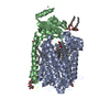
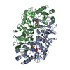
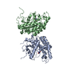
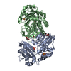
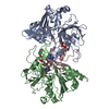
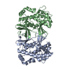
- PDB-3csm: STRUCTURE OF YEAST CHORISMATE MUTASE WITH BOUND TRP AND AN ENDOOX... -
+ Open data

- Basic information
| Entry | Database: PDB / ID: 3csm | ||||||
|---|---|---|---|---|---|---|---|
| Title | STRUCTURE OF YEAST CHORISMATE MUTASE WITH BOUND TRP AND AN ENDOOXABICYCLIC INHIBITOR | ||||||
 Components Components | CHORISMATE MUTASE | ||||||
 Keywords Keywords | COMPLEX (ISOMERASE/PEPTIDE) / CHORISMATE PYRUVATEMUTASE / ALLOSTERIC PROTEIN / COMPLEX (ISOMERASE-PEPTIDE) / TRANSITION STATE ANALOG / COMPLEX (ISOMERASE-PEPTIDE) complex | ||||||
| Function / homology |  Function and homology information Function and homology informationtryptophan binding / L-tyrosine binding / L-tyrosine biosynthetic process / chorismate metabolic process / chorismate mutase / chorismate mutase activity / L-phenylalanine biosynthetic process / aromatic amino acid biosynthetic process / nucleus / cytoplasm Similarity search - Function | ||||||
| Biological species |  | ||||||
| Method |  X-RAY DIFFRACTION / MOLECULAR REPLACEMENT / Resolution: 3 Å X-RAY DIFFRACTION / MOLECULAR REPLACEMENT / Resolution: 3 Å | ||||||
 Authors Authors | Straeter, N. / Schnappauf, G. / Braus, G. / Lipscomb, W.N. | ||||||
 Citation Citation | Journal: Structure / Year: 1997 Title: Mechanisms of catalysis and allosteric regulation of yeast chorismate mutase from crystal structures. Authors: Strater, N. / Schnappauf, G. / Braus, G. / Lipscomb, W.N. #1: Journal: Proc.Natl.Acad.Sci.USA / Year: 1996Title: Crystal Structure of the T State of Allosteric Yeast Chorismate Mutase and Comparison with the R State Authors: Strater, N. / Hakansson, K. / Schnappauf, G. / Braus, G. / Lipscomb, W.N. #2: Journal: Proc.Natl.Acad.Sci.USA / Year: 1995Title: Location of the Active Site of Allosteric Chorismate Mutase from Saccharomyces Cerevisiae, and Comments on the Catalytic and Regulatory Mechanisms Authors: Xue, Y. / Lipscomb, W.N. #3: Journal: Proc.Natl.Acad.Sci.USA / Year: 1994Title: The Crystal Structure of Allosteric Chorismate Mutase at 2.2-A Resolution Authors: Xue, Y. / Lipscomb, W.N. / Graf, R. / Schnappauf, G. / Braus, G. | ||||||
| History |
|
- Structure visualization
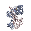
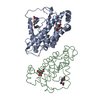
| Structure viewer | Molecule: MolmilJmol/JSmol |
|---|
- Downloads & links
-Download
| PDBx/mmCIF format | 3csm.cif.gz | 113.2 KB | Display | PDBx/mmCIF format |
|---|---|---|---|---|
| PDB format | pdb3csm.ent.gz | 89.6 KB | Display | PDB format |
| PDBx/mmJSON format | 3csm.json.gz | Tree view | PDBx/mmJSON format | |
| Others |  Other downloads Other downloads |
-Validation report
| Arichive directory | https://data.pdbj.org/pub/pdb/validation_reports/cs/3csmftp://data.pdbj.org/pub/pdb/validation_reports/cs/3csm | HTTPS FTP |
|---|
-Related structure data
| Related structure data |  4csmC 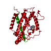 5csmC 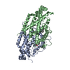 1csmS S: Starting model for refinement C: citing same article ( |
|---|---|
| Similar structure data |
-Links
 PDBj
PDBj

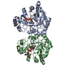
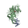
- Assembly
| Deposited unit | 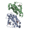
| ||||||||
|---|---|---|---|---|---|---|---|---|---|
| 1 |
| ||||||||
| 2 | 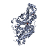
| ||||||||
| 3 | 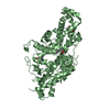
| ||||||||
| Unit cell |
|
-Components
| #1: Protein | Mass: 29958.484 Da / Num. of mol.: 2 Source method: isolated from a genetically manipulated source Source: (gene. exp.) Strain: RH1242 / Plasmid: PME605 / Production host: #2: Chemical |   Type: L-peptide linking / Mass: 204.225 Da / Num. of mol.: 2 / Source method: obtained synthetically / Formula: C11H12N2O2 / Details: TRYPTOPHAN BOUND TO A REGULATORY SITE Type: L-peptide linking / Mass: 204.225 Da / Num. of mol.: 2 / Source method: obtained synthetically / Formula: C11H12N2O2 / Details: TRYPTOPHAN BOUND TO A REGULATORY SITE#3: Chemical |   Mass: 228.199 Da / Num. of mol.: 2 / Source method: obtained synthetically / Formula: C10H12O6 Mass: 228.199 Da / Num. of mol.: 2 / Source method: obtained synthetically / Formula: C10H12O6 |
|---|
-Experimental details
-Experiment
| Experiment | Method: X-RAY DIFFRACTION / Number of used crystals: 1 |
|---|
- Sample preparation
| Crystal | Density Matthews: 4.3 Å3/Da / Density % sol: 71.24 % | ||||||||||||||||||||||||||||||||||||||||
|---|---|---|---|---|---|---|---|---|---|---|---|---|---|---|---|---|---|---|---|---|---|---|---|---|---|---|---|---|---|---|---|---|---|---|---|---|---|---|---|---|---|
| Crystal grow | Method: vapor diffusion, hanging drop / pH: 8 Details: HANGING DROP, 19 % (W/V) PEG MONOMETHYLETHER 5000, 5 MM DTT, 0.15 M TRIS PH 8.0, 0.15 M SODIUM ACETATE, 16 MM TRYPTOPHAN, 3 MM INHIBITOR, 10 MG/ML PROTEIN, vapor diffusion - hanging drop | ||||||||||||||||||||||||||||||||||||||||
| Crystal grow | *PLUS Method: vapor diffusion, hanging drop | ||||||||||||||||||||||||||||||||||||||||
| Components of the solutions | *PLUS
|
-Data collection
| Diffraction | Mean temperature: 123 K |
|---|---|
| Diffraction source | Source: ROTATING ANODE / Type: ELLIOTT GX-13 / Wavelength: 1.5418 |
| Detector | Type: SIEMENS-NICOLET X100 / Detector: AREA DETECTOR / Date: Oct 15, 1996 / Details: SUPPER DOUBLE-MIRRORS |
| Radiation | Monochromator: DOUBLE CRYSTAL SI(111) / Monochromatic (M) / Laue (L): M / Scattering type: x-ray |
| Radiation wavelength | Wavelength: 1.5418 Å / Relative weight: 1 |
| Reflection | Resolution: 3→20 Å / Num. obs: 20253 / % possible obs: 98.4 % / Observed criterion σ(I): 0 / Redundancy: 2.5 % / Rmerge(I) obs: 0.092 |
| Reflection shell | Resolution: 3→3.2 Å / Rmerge(I) obs: 0.325 / % possible all: 99.1 |
| Reflection | *PLUS Num. measured all: 51388 |
| Reflection shell | *PLUS % possible obs: 99.1 % |
- Processing
| Software |
| ||||||||||||||||||||||||||||||||||||||||||||||||||||||||||||||||||||||||||||||||
|---|---|---|---|---|---|---|---|---|---|---|---|---|---|---|---|---|---|---|---|---|---|---|---|---|---|---|---|---|---|---|---|---|---|---|---|---|---|---|---|---|---|---|---|---|---|---|---|---|---|---|---|---|---|---|---|---|---|---|---|---|---|---|---|---|---|---|---|---|---|---|---|---|---|---|---|---|---|---|---|---|---|
| Refinement | Method to determine structure: MOLECULAR REPLACEMENT Starting model: PDB ENTRY 1CSM Resolution: 3→15 Å / σ(F): 2
| ||||||||||||||||||||||||||||||||||||||||||||||||||||||||||||||||||||||||||||||||
| Displacement parameters | Biso mean: 38.6 Å2 | ||||||||||||||||||||||||||||||||||||||||||||||||||||||||||||||||||||||||||||||||
| Refinement step | Cycle: LAST / Resolution: 3→15 Å
| ||||||||||||||||||||||||||||||||||||||||||||||||||||||||||||||||||||||||||||||||
| Refine LS restraints |
| ||||||||||||||||||||||||||||||||||||||||||||||||||||||||||||||||||||||||||||||||
| Refine LS restraints NCS | NCS model details: CONSTRAINTS | ||||||||||||||||||||||||||||||||||||||||||||||||||||||||||||||||||||||||||||||||
| LS refinement shell | Resolution: 3→3.14 Å / Total num. of bins used: 8
| ||||||||||||||||||||||||||||||||||||||||||||||||||||||||||||||||||||||||||||||||
| Xplor file |
| ||||||||||||||||||||||||||||||||||||||||||||||||||||||||||||||||||||||||||||||||
| Software | *PLUS Name: X-PLOR / Version: 3.851 / Classification: refinement | ||||||||||||||||||||||||||||||||||||||||||||||||||||||||||||||||||||||||||||||||
| Refinement | *PLUS | ||||||||||||||||||||||||||||||||||||||||||||||||||||||||||||||||||||||||||||||||
| Solvent computation | *PLUS | ||||||||||||||||||||||||||||||||||||||||||||||||||||||||||||||||||||||||||||||||
| Displacement parameters | *PLUS | ||||||||||||||||||||||||||||||||||||||||||||||||||||||||||||||||||||||||||||||||
| Refine LS restraints | *PLUS
| ||||||||||||||||||||||||||||||||||||||||||||||||||||||||||||||||||||||||||||||||
| LS refinement shell | *PLUS Lowest resolution: 3.2 Å / Rfactor obs: 0.285 |
