 Movie
Movie Controller


+ Open data

- Basic information
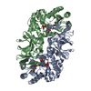
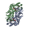
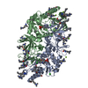
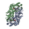
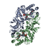
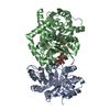
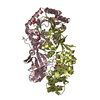
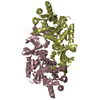
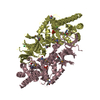
| Entry | Database: PDB / ID: 1bd0 | ||||||
|---|---|---|---|---|---|---|---|
| Title | ALANINE RACEMASE COMPLEXED WITH ALANINE PHOSPHONATE | ||||||
 Components Components | ALANINE RACEMASE | ||||||
 Keywords Keywords | ISOMERASE / ALANINE / PYRIDOXAL PHOSPHATE / ALANINE PHOSPHONATE | ||||||
| Function / homology |  Function and homology information Function and homology informationalanine racemase / D-alanine biosynthetic process / alanine racemase activity / peptidoglycan biosynthetic process / pyridoxal phosphate binding / cytosol Similarity search - Function | ||||||
| Biological species |   Geobacillus stearothermophilus (bacteria) Geobacillus stearothermophilus (bacteria) | ||||||
| Method |  X-RAY DIFFRACTION / SYNCHROTRON / MOLECULAR REPLACEMENT / Resolution: 1.6 Å X-RAY DIFFRACTION / SYNCHROTRON / MOLECULAR REPLACEMENT / Resolution: 1.6 Å | ||||||
 Authors Authors | Stamper, G.F. / Morollo, A.A. / Ringe, D. | ||||||
 Citation Citation | Journal: Biochemistry / Year: 1998 Title: Reaction of alanine racemase with 1-aminoethylphosphonic acid forms a stable external aldimine. Authors: Stamper, G.F. / Morollo, A.A. / Ringe, D. / Stamper, C.G. #1: Journal: Biochemistry / Year: 1997Title: Determination of the Structure of Alanine Racemase from Bacillus Stearothermophilus at 1.9-A Resolution Authors: Shaw, J.P. / Petsko, G.A. / Ringe, D. #2: Journal: J.Biol.Chem. / Year: 1987Title: X-Ray Crystallographic Studies of the Alanine-Specific Racemase from Bacillus Stearothermophilus. Overproduction, Crystallization, and Preliminary Characterization Authors: Neidhart, D.J. / Distefano, M.D. / Tanizawa, K. / Soda, K. / Walsh, C.T. / Petsko, G.A. | ||||||
| History |
|
- Structure visualization
| Structure viewer | Molecule: MolmilJmol/JSmol |
|---|
- Downloads & links
-Download
| PDBx/mmCIF format | 1bd0.cif.gz | 167 KB | Display | PDBx/mmCIF format |
|---|---|---|---|---|
| PDB format | pdb1bd0.ent.gz | 133.1 KB | Display | PDB format |
| PDBx/mmJSON format | 1bd0.json.gz | Tree view | PDBx/mmJSON format | |
| Others |  Other downloads Other downloads |
-Validation report
| Arichive directory | https://data.pdbj.org/pub/pdb/validation_reports/bd/1bd0ftp://data.pdbj.org/pub/pdb/validation_reports/bd/1bd0 | HTTPS FTP |
|---|
-Related structure data
| Related structure data |  1sftS S: Starting model for refinement |
|---|---|
| Similar structure data |

-Links
 PDBj
PDBj- Assembly
| Deposited unit | 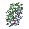
| ||||||||
|---|---|---|---|---|---|---|---|---|---|
| 1 |
| ||||||||
| Unit cell |
|
-Components
| #1: Protein | Mass: 43565.953 Da / Num. of mol.: 2 / Source method: isolated from a natural source / Source: (natural) Geobacillus stearothermophilus (bacteria) / Plasmid: PMDALR3 / Strain: XL-1 BLUE / References: UniProt: P10724, alanine racemase#2: Chemical | 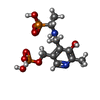  Mass: 356.206 Da / Num. of mol.: 2 / Source method: obtained synthetically / Formula: C10H18N2O8P2 Mass: 356.206 Da / Num. of mol.: 2 / Source method: obtained synthetically / Formula: C10H18N2O8P2#3: Water | ChemComp-HOH / |  Mass: 18.015 Da / Num. of mol.: 387 / Source method: isolated from a natural source / Formula: H2O Mass: 18.015 Da / Num. of mol.: 387 / Source method: isolated from a natural source / Formula: H2O |
|---|
-Experimental details
-Experiment
| Experiment | Method: X-RAY DIFFRACTION / Number of used crystals: 1 |
|---|
- Sample preparation
| Crystal | Density Matthews: 2.12 Å3/Da / Density % sol: 41.5 % | ||||||||||||||||||||||||||||||||||||
|---|---|---|---|---|---|---|---|---|---|---|---|---|---|---|---|---|---|---|---|---|---|---|---|---|---|---|---|---|---|---|---|---|---|---|---|---|---|
| Crystal grow | pH: 8.5 Details: PROTEIN WAS CRYSTALLIZED FROM 100MM TRIS BUFFER, PH 8.5, 200MM SODIUM ACETATE, 21% PEG 4000, AND 4MM 1-AMINOETHYL PHOSPHONIC ACID. | ||||||||||||||||||||||||||||||||||||
| Crystal | *PLUS | ||||||||||||||||||||||||||||||||||||
| Crystal grow | *PLUS Method: vapor diffusion, hanging drop | ||||||||||||||||||||||||||||||||||||
| Components of the solutions | *PLUS
|
-Data collection
| Diffraction | Mean temperature: 95 K |
|---|---|
| Diffraction source | Source: SYNCHROTRON / Site: NSLS  / Beamline: X12C / Wavelength: 1.15 / Beamline: X12C / Wavelength: 1.15 |
| Detector | Type: BRANDEIS / Detector: CCD / Date: Oct 30, 1996 / Details: MIRROR |
| Radiation | Monochromator: SI(111) / Monochromatic (M) / Laue (L): M / Scattering type: x-ray |
| Radiation wavelength | Wavelength: 1.15 Å / Relative weight: 1 |
| Reflection | Resolution: 1.6→25 Å / Num. obs: 97731 / % possible obs: 96.7 % / Observed criterion σ(I): 0 / Redundancy: 5.2 % / Rmerge(I) obs: 0.078 / Rsym value: 0.078 / Net I/σ(I): 12.1 |
| Reflection shell | Resolution: 1.6→1.66 Å / Redundancy: 2.2 % / Mean I/σ(I) obs: 0.8 / % possible all: 93.7 |
| Reflection | *PLUS Num. measured all: 516378 / Rmerge F obs: 0.078 |
| Reflection shell | *PLUS % possible obs: 93.7 % |
- Processing
| Software |
| ||||||||||||||||||||||||||||||||||||||||||||||||||||||||||||
|---|---|---|---|---|---|---|---|---|---|---|---|---|---|---|---|---|---|---|---|---|---|---|---|---|---|---|---|---|---|---|---|---|---|---|---|---|---|---|---|---|---|---|---|---|---|---|---|---|---|---|---|---|---|---|---|---|---|---|---|---|---|
| Refinement | Method to determine structure: MOLECULAR REPLACEMENT Starting model: PDB ENTRY 1SFT Resolution: 1.6→10 Å / Rfactor Rfree error: 0.005 / Data cutoff high absF: 10000 / Data cutoff low absF: 0.001 / Cross valid method: THROUGHOUT / σ(F): 0
| ||||||||||||||||||||||||||||||||||||||||||||||||||||||||||||
| Refinement step | Cycle: LAST / Resolution: 1.6→10 Å
| ||||||||||||||||||||||||||||||||||||||||||||||||||||||||||||
| Refine LS restraints |
| ||||||||||||||||||||||||||||||||||||||||||||||||||||||||||||
| Xplor file |
| ||||||||||||||||||||||||||||||||||||||||||||||||||||||||||||
| Software | *PLUS Name: X-PLOR / Version: 3.851 / Classification: refinement | ||||||||||||||||||||||||||||||||||||||||||||||||||||||||||||
| Refinement | *PLUS Rfactor obs: 0.24 / Rfactor Rwork: 0.24 | ||||||||||||||||||||||||||||||||||||||||||||||||||||||||||||
| Solvent computation | *PLUS | ||||||||||||||||||||||||||||||||||||||||||||||||||||||||||||
| Displacement parameters | *PLUS | ||||||||||||||||||||||||||||||||||||||||||||||||||||||||||||
| Refine LS restraints | *PLUS
|
