 Movie
Movie Controller

[English] 日本語
 Yorodumi
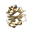
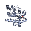
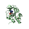
- PDB-3cr6: Crystal Structure of the R132K:R111L:A32E Mutant of Cellular Reti... -
+ Open data

- Basic information
| Entry | Database: PDB / ID: 3cr6 | ||||||
|---|---|---|---|---|---|---|---|
| Title | Crystal Structure of the R132K:R111L:A32E Mutant of Cellular Retinoic Acid Binding Protein Type II Complexed with C15-aldehyde (a retinal analog) at 1.22 Angstrom resolution. | ||||||
 Components Components | Cellular retinoic acid-binding protein 2 | ||||||
 Keywords Keywords | TRANSPORT PROTEIN / CRABPII / retinal / schiff base / protonated schiff base / PSB / C15-aldehyde / retinoic acid / retinoid / Nucleus / Retinol-binding / Transport / Vitamin A | ||||||
| Function / homology |  Function and homology information Function and homology informationpositive regulation of collateral sprouting / retinoid binding / retinoic acid binding / retinal binding / embryonic forelimb morphogenesis / retinoic acid metabolic process / retinol binding / Signaling by Retinoic Acid / epidermis development / fatty acid transport ...positive regulation of collateral sprouting / retinoid binding / retinoic acid binding / retinal binding / embryonic forelimb morphogenesis / retinoic acid metabolic process / retinol binding / Signaling by Retinoic Acid / epidermis development / fatty acid transport / cyclin binding / fatty acid binding / regulation of DNA-templated transcription / endoplasmic reticulum / signal transduction / extracellular exosome / nucleoplasm / nucleus / cytoplasm / cytosol Similarity search - Function | ||||||
| Biological species |  Homo sapiens (human) Homo sapiens (human) | ||||||
| Method |  X-RAY DIFFRACTION / SYNCHROTRON / MOLECULAR REPLACEMENT / molecular replacement / Resolution: 1.22 Å X-RAY DIFFRACTION / SYNCHROTRON / MOLECULAR REPLACEMENT / molecular replacement / Resolution: 1.22 Å | ||||||
 Authors Authors | Jia, X. / Geiger, J.H. | ||||||
 Citation Citation | Journal: To be Published Title: Two distinctive orientations of binding determined by a single mutation in the CRABPII mutant-C15-aldehyde complexes Authors: Jia, X. / Lee, K.S. / Vasileiou, C. / Borhan, B. / Geiger, J.H. #1: Journal: To be PublishedTitle: Protein engineering: wavelength regulation mechanism investigated in a rhodopsin mimic Authors: Lee, K.S. / Jia, X. / Vasileiou, C. / Geiger, J.H. / Borhan, B. | ||||||
| History |
|
- Structure visualization
| Structure viewer | Molecule: MolmilJmol/JSmol |
|---|
- Downloads & links
-Download
| PDBx/mmCIF format | 3cr6.cif.gz | 50.8 KB | Display | PDBx/mmCIF format |
|---|---|---|---|---|
| PDB format | pdb3cr6.ent.gz | 34.2 KB | Display | PDB format |
| PDBx/mmJSON format | 3cr6.json.gz | Tree view | PDBx/mmJSON format | |
| Others |  Other downloads Other downloads |
-Validation report
| Arichive directory | https://data.pdbj.org/pub/pdb/validation_reports/cr/3cr6ftp://data.pdbj.org/pub/pdb/validation_reports/cr/3cr6 | HTTPS FTP |
|---|
-Related structure data
| Related structure data |  3f9dC  3fa6C 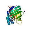 2g7bS S: Starting model for refinement C: citing same article ( |
|---|---|
| Similar structure data |


-Links
 PDBj
PDBj


- Assembly
| Deposited unit | 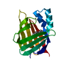
| ||||||||
|---|---|---|---|---|---|---|---|---|---|
| 1 |
| ||||||||
| Unit cell |
|
-Components
| #1: Protein | Mass: 15567.790 Da / Num. of mol.: 1 / Mutation: R132K, R111L, A32E Source method: isolated from a genetically manipulated source Source: (gene. exp.) Homo sapiens (human) / Gene: CRABP2 / Plasmid: CRABPII-pET17b-KL-A32E / Production host:  |
|---|---|
| #2: Chemical | ChemComp-LSR / 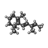  Mass: 204.351 Da / Num. of mol.: 1 / Source method: obtained synthetically / Formula: C15H24 Mass: 204.351 Da / Num. of mol.: 1 / Source method: obtained synthetically / Formula: C15H24 |
| #3: Water | ChemComp-HOH /  Mass: 18.015 Da / Num. of mol.: 310 / Source method: isolated from a natural source / Formula: H2O Mass: 18.015 Da / Num. of mol.: 310 / Source method: isolated from a natural source / Formula: H2O |
| Has protein modification | Y |
-Experimental details
-Experiment
| Experiment | Method: X-RAY DIFFRACTION / Number of used crystals: 1 |
|---|
- Sample preparation
| Crystal | Density Matthews: 1.93 Å3/Da / Density % sol: 36.42 % |
|---|---|
| Crystal grow | Temperature: 277 K / Method: vapor diffusion, hanging drop / pH: 7.5 Details: 0.1M sodium citrate, 0.2M ammonium acetate, 26% PEG 4000, pH 7.5, VAPOR DIFFUSION, HANGING DROP, temperature 277K |
-Data collection
| Diffraction | Mean temperature: 100 K |
|---|---|
| Diffraction source | Source: SYNCHROTRON / Site: APS  / Beamline: 21-ID-D / Wavelength: 0.97869 Å / Beamline: 21-ID-D / Wavelength: 0.97869 Å |
| Detector | Type: MARMOSAIC 300 mm CCD / Detector: CCD / Date: Apr 8, 2007 |
| Radiation | Monochromator: C(111) / Protocol: SINGLE WAVELENGTH / Monochromatic (M) / Laue (L): M / Scattering type: x-ray |
| Radiation wavelength | Wavelength: 0.97869 Å / Relative weight: 1 |
| Reflection | Resolution: 1.03→50 Å / Num. obs: 42691 / % possible obs: 72.4 % / Observed criterion σ(I): 1 / Redundancy: 3.8 % / Biso Wilson estimate: 14.5 Å2 / Rmerge(I) obs: 0.046 / Χ2: 0.982 / Net I/σ(I): 9.5 |
| Reflection shell | Resolution: 1.03→1.07 Å / Redundancy: 1.4 % / Rmerge(I) obs: 0.806 / Mean I/σ(I) obs: 0.759 / Num. unique all: 284 / Χ2: 0.687 / % possible all: 4.8 |
-Phasing
| Phasing | Method: molecular replacement | |||||||||
|---|---|---|---|---|---|---|---|---|---|---|
| Phasing MR | Rfactor: 0.533 / Cor.coef. Fo:Fc: 0.457
|
- Processing
| Software |
| ||||||||||||||||||||||||||||||||||||||||
|---|---|---|---|---|---|---|---|---|---|---|---|---|---|---|---|---|---|---|---|---|---|---|---|---|---|---|---|---|---|---|---|---|---|---|---|---|---|---|---|---|---|
| Refinement | Method to determine structure: MOLECULAR REPLACEMENT Starting model: PDB entry 2G7B Resolution: 1.22→10 Å / σ(F): 4
| ||||||||||||||||||||||||||||||||||||||||
| Displacement parameters | Biso mean: 20.928 Å2 | ||||||||||||||||||||||||||||||||||||||||
| Refinement step | Cycle: LAST / Resolution: 1.22→10 Å
| ||||||||||||||||||||||||||||||||||||||||
| Refine LS restraints |
| ||||||||||||||||||||||||||||||||||||||||
| LS refinement shell | Resolution: 1.22→1.252 Å
|
