 Movie
Movie Controller

[English] 日本語
 Yorodumi
- PDB-7jsl: Crystal structure of the DNA binding domain of human transcriptio... -
+ Open data

- Basic information
| Entry | Database: PDB / ID: 7jsl | |||||||||
|---|---|---|---|---|---|---|---|---|---|---|
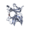
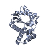
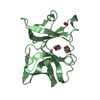
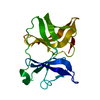
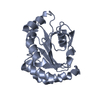
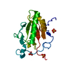
| Title | Crystal structure of the DNA binding domain of human transcription factor ERF in the oxidized form, in complex with double-stranded DNA ACCGGAAGTG | |||||||||
 Components Components |
| |||||||||
 Keywords Keywords | DNA BINDING PROTEIN/DNA / Transcription / Tumor suppressor / ETS family / repressor / DNA BINDING PROTEIN / DNA BINDING PROTEIN-DNA complex | |||||||||
| Function / homology |  Function and homology information Function and homology informationOncogene Induced Senescence / DNA-binding transcription repressor activity, RNA polymerase II-specific / sequence-specific DNA binding / DNA-binding transcription factor activity, RNA polymerase II-specific / cell differentiation / regulation of transcription by RNA polymerase II / chromatin / negative regulation of transcription by RNA polymerase II / nucleoplasm / nucleus ...Oncogene Induced Senescence / DNA-binding transcription repressor activity, RNA polymerase II-specific / sequence-specific DNA binding / DNA-binding transcription factor activity, RNA polymerase II-specific / cell differentiation / regulation of transcription by RNA polymerase II / chromatin / negative regulation of transcription by RNA polymerase II / nucleoplasm / nucleus / plasma membrane / cytosol Similarity search - Function | |||||||||
| Biological species |  Homo sapiens (human) Homo sapiens (human)synthetic construct (others) | |||||||||
| Method |  X-RAY DIFFRACTION / SYNCHROTRON / MOLECULAR REPLACEMENT / Resolution: 4.51 Å X-RAY DIFFRACTION / SYNCHROTRON / MOLECULAR REPLACEMENT / Resolution: 4.51 Å | |||||||||
 Authors Authors | Hou, C. / Tsodikov, O.V. | |||||||||
| Funding support |  United States, 2items United States, 2items
| |||||||||
 Citation Citation | Journal: Biochemistry / Year: 2020 Title: Structural Insight into the DNA Binding Function of Transcription Factor ERF. Authors: Hou, C. / McCown, C. / Ivanov, D.N. / Tsodikov, O.V. | |||||||||
| History |
|
- Structure visualization
| Structure viewer | Molecule: MolmilJmol/JSmol |
|---|
- Downloads & links
-Download
| PDBx/mmCIF format | 7jsl.cif.gz | 129.8 KB | Display | PDBx/mmCIF format |
|---|---|---|---|---|
| PDB format | pdb7jsl.ent.gz | 96.6 KB | Display | PDB format |
| PDBx/mmJSON format | 7jsl.json.gz | Tree view | PDBx/mmJSON format | |
| Others |  Other downloads Other downloads |
-Validation report
| Arichive directory | https://data.pdbj.org/pub/pdb/validation_reports/js/7jslftp://data.pdbj.org/pub/pdb/validation_reports/js/7jsl | HTTPS FTP |
|---|
-Related structure data
| Related structure data |  7jsaSC S: Starting model for refinement C: citing same article ( |
|---|---|
| Similar structure data |


-Links
 PDBj
PDBj

- Assembly
| Deposited unit | 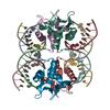
| ||||||||
|---|---|---|---|---|---|---|---|---|---|
| 1 | 
| ||||||||
| 2 | 
| ||||||||
| 3 | 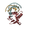
| ||||||||
| 4 | 
| ||||||||
| Unit cell |
|
-Components
| #1: DNA chain | Mass: 3094.042 Da / Num. of mol.: 4 / Source method: obtained synthetically / Details: synthetic DNA / Source: (synth.) synthetic construct (others) #2: DNA chain | Mass: 2995.967 Da / Num. of mol.: 4 / Source method: obtained synthetically / Details: synthetic DNA / Source: (synth.) synthetic construct (others) #3: Protein | Mass: 14222.460 Da / Num. of mol.: 4 Source method: isolated from a genetically manipulated source Details: The N-terminal region GPHM is a leftover after affinity tag cleavage. The C-terminal region KLVL...SGSS is disordered. Source: (gene. exp.) Homo sapiens (human) / Gene: ERF / Production host:  Has protein modification | Y | |
|---|
-Experimental details
-Experiment
| Experiment | Method: X-RAY DIFFRACTION / Number of used crystals: 1 |
|---|
- Sample preparation
| Crystal | Density Matthews: 4.41 Å3/Da / Density % sol: 72.1 % |
|---|---|
| Crystal grow | Temperature: 294 K / Method: vapor diffusion, hanging drop / pH: 7.5 / Details: 10% PEG 4000, 0.1M Hepes pH 7.5 |
-Data collection
| Diffraction | Mean temperature: 100 K / Serial crystal experiment: N |
|---|---|
| Diffraction source | Source: SYNCHROTRON / Site: APS / Beamline: 21-ID-D / Wavelength: 1 Å |
| Detector | Type: DECTRIS EIGER X 16M / Detector: PIXEL / Date: Oct 8, 2017 |
| Radiation | Protocol: SINGLE WAVELENGTH / Monochromatic (M) / Laue (L): M / Scattering type: x-ray |
| Radiation wavelength | Wavelength: 1 Å / Relative weight: 1 |
| Reflection | Resolution: 4.5→50 Å / Num. obs: 8678 / % possible obs: 99.5 % / Redundancy: 5.7 % / Rmerge(I) obs: 0.082 / Net I/σ(I): 17.9 |
| Reflection shell | Resolution: 4.5→4.58 Å / Num. unique obs: 418 / CC1/2: 0.73 |
- Processing
| Software |
| ||||||||||||||||||||||||||||||||||||||||||||||||||||||||||||
|---|---|---|---|---|---|---|---|---|---|---|---|---|---|---|---|---|---|---|---|---|---|---|---|---|---|---|---|---|---|---|---|---|---|---|---|---|---|---|---|---|---|---|---|---|---|---|---|---|---|---|---|---|---|---|---|---|---|---|---|---|---|
| Refinement | Method to determine structure: MOLECULAR REPLACEMENT Starting model: 7JSA Resolution: 4.51→36.85 Å / Cor.coef. Fo:Fc: 0.917 / Cor.coef. Fo:Fc free: 0.946 / SU B: 71.617 / SU ML: 0.784 / Cross valid method: THROUGHOUT / σ(F): 0 / ESU R Free: 0.899 / Stereochemistry target values: MAXIMUM LIKELIHOOD Details: HYDROGENS HAVE BEEN ADDED IN THE RIDING POSITIONS U VALUES : REFINED INDIVIDUALLY
| ||||||||||||||||||||||||||||||||||||||||||||||||||||||||||||
| Solvent computation | Ion probe radii: 0.8 Å / Shrinkage radii: 0.8 Å / VDW probe radii: 1.2 Å / Solvent model: MASK | ||||||||||||||||||||||||||||||||||||||||||||||||||||||||||||
| Displacement parameters | Biso max: 347.29 Å2 / Biso mean: 212.875 Å2 / Biso min: 138.21 Å2
| ||||||||||||||||||||||||||||||||||||||||||||||||||||||||||||
| Refinement step | Cycle: final / Resolution: 4.51→36.85 Å
| ||||||||||||||||||||||||||||||||||||||||||||||||||||||||||||
| Refine LS restraints |
| ||||||||||||||||||||||||||||||||||||||||||||||||||||||||||||
| LS refinement shell | Resolution: 4.512→4.628 Å / Rfactor Rfree error: 0 / Total num. of bins used: 20
|
