 Yorodumi
+ Open data
- Basic information
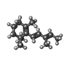
| Entry |  Database: PDB chemical components / ID: LSR Database: PDB chemical components / ID: LSR |
|---|---|
| Name | Name: |
-Chemical information
| Composition |  | ||||
|---|---|---|---|---|---|
| Others | Type: NON-POLYMER / PDB classification: HETAIN / Three letter code: LSR / Ideal coordinates details: Corina / Model coordinates PDB-ID: 3CR6 | ||||
| History |
| ||||
 External links External links |  UniChem / UniChem /  ChemSpider / ChemSpider /  DrugBank / DrugBank /  Nikkaji / Nikkaji /  PubChem / PubChem /  SureChEMBL / SureChEMBL /  ZINC / ZINC /  Wikipedia search / Wikipedia search /  Google search Google search |
- Structure visualization
| Structure viewer | Molecule:  Molmil Molmil Jmol/JSmol Jmol/JSmol |
|---|
-Details
-SMILES
| ACDLabs 10.04 | | CACTVS 3.341 | OpenEye OEToolkits 1.5.0 | |
|---|
-SMILES CANONICAL
| CACTVS 3.341 | | OpenEye OEToolkits 1.5.0 | |
|---|
-InChI
| InChI 1.03 |
|---|
-InChIKey
| InChI 1.03 |
|---|
-SYSTEMATIC NAME
| ACDLabs 10.04 | | OpenEye OEToolkits 1.5.0 | |
|---|
-PDB entries
Showing all 4 items

PDB-3cr6: 
Crystal Structure of the R132K:R111L:A32E Mutant of Cellular Retinoic Acid Binding Protein Type II Complexed with C15-aldehyde (a retinal analog) at 1.22 Angstrom resolution.

PDB-3f8a: 
Crystal Structure of the R132K:R111L:L121E:R59W Mutant of Cellular Retinoic Acid-Binding Protein Type II Complexed with C15-aldehyde (a retinal analog) at 1.95 Angstrom resolution.

PDB-3f9d: 
Crystal structure of the R132K:R111L:T54E mutant of cellular retinoic acid-binding protein II complexed with C15-aldehyde (a retinal analog) at 2.00 angstrom resolution

PDB-3fa6: 
Crystal structure of the R132K:Y134F:R111L:L121D:T54V mutant of cellular retinoic acid-binding protein II complexed with C15-aldehyde (a retinal analog) at 1.54 angstrom resolution
 Movie
Movie Controller


