 Movie
Movie Controller

[English] 日本語
 Yorodumi
- PDB-1oqv: Structure of TcpA, the Type IV pilin subunit from the toxin co-re... -
+ Open data

- Basic information
| Entry | Database: PDB / ID: 1oqv | ||||||
|---|---|---|---|---|---|---|---|
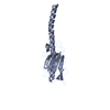
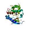
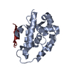
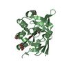
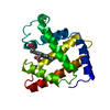
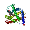
| Title | Structure of TcpA, the Type IV pilin subunit from the toxin co-regulated pilus of Vibrio cholerae classical biotype | ||||||
 Components Components | toxin-coregulated pilus subunit | ||||||
 Keywords Keywords | CELL ADHESION / TcpA / Type IV pilin / pilin / pilus filament / Vibrio cholerae / fiber forming protein / adhesin / fimbriae | ||||||
| Function / homology |  Function and homology information Function and homology information | ||||||
| Biological species |   Vibrio cholerae (bacteria) Vibrio cholerae (bacteria) | ||||||
| Method |  X-RAY DIFFRACTION / SYNCHROTRON / MOLECULAR REPLACEMENT / Resolution: 1.3 Å X-RAY DIFFRACTION / SYNCHROTRON / MOLECULAR REPLACEMENT / Resolution: 1.3 Å | ||||||
 Authors Authors | Craig, L. / Tainer, J.A. | ||||||
 Citation Citation | Journal: Mol.Cell / Year: 2003 Title: Type IV Pilin Structure and Assembly: X-Ray and EM Analyses of Vibrio cholerae Toxin-Coregulated Pilus and Pseudomonas aeruginosa PAK Pilin Authors: Craig, L. / Taylor, R.K. / Pique, M.E. / Adair, B.D. / Arvai, A.S. / Singh, M. / Lloyd, S.J. / Shin, D.S. / Getzoff, E.D. / Yeager, M. / Forest, K.T. / Tainer, J.A. | ||||||
| History |
|
- Structure visualization
| Structure viewer | Molecule: MolmilJmol/JSmol |
|---|
- Downloads & links
-Download
| PDBx/mmCIF format | 1oqv.cif.gz | 127.1 KB | Display | PDBx/mmCIF format |
|---|---|---|---|---|
| PDB format | pdb1oqv.ent.gz | 96.9 KB | Display | PDB format |
| PDBx/mmJSON format | 1oqv.json.gz | Tree view | PDBx/mmJSON format | |
| Others |  Other downloads Other downloads |
-Validation report
| Arichive directory | https://data.pdbj.org/pub/pdb/validation_reports/oq/1oqvftp://data.pdbj.org/pub/pdb/validation_reports/oq/1oqv | HTTPS FTP |
|---|
-Related structure data


-Links
 PDBj
PDBj
- Assembly
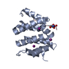
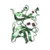
| Deposited unit | 
| ||||||||
|---|---|---|---|---|---|---|---|---|---|
| 1 | 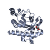
| ||||||||
| 2 | 
| ||||||||
| 3 | 
| ||||||||
| Unit cell |
| ||||||||
| Details | thousands of pilin subunits assemble to form a long thin filament 6 nm in diameter and several microns in length (see TCP model, PDB file XXX) |
-Components
| #1: Protein | Mass: 19794.285 Da / Num. of mol.: 3 / Fragment: globular head domain, residues 29-199 Source method: isolated from a genetically manipulated source Source: (gene. exp.) Vibrio cholerae (bacteria) / Gene: tcpa / Plasmid: pET-15b / Production host: #2: Chemical |   Mass: 92.094 Da / Num. of mol.: 3 / Source method: obtained synthetically / Formula: C3H8O3 Mass: 92.094 Da / Num. of mol.: 3 / Source method: obtained synthetically / Formula: C3H8O3#3: Water | ChemComp-HOH / |  Mass: 18.015 Da / Num. of mol.: 765 / Source method: isolated from a natural source / Formula: H2O Mass: 18.015 Da / Num. of mol.: 765 / Source method: isolated from a natural source / Formula: H2OHas protein modification | Y | |
|---|
-Experimental details
-Experiment
| Experiment | Method: X-RAY DIFFRACTION / Number of used crystals: 1 |
|---|
- Sample preparation
| Crystal | Density Matthews: 1.99 Å3/Da / Density % sol: 37.74 % | ||||||||||||||||||||||||||||||||||||||||||
|---|---|---|---|---|---|---|---|---|---|---|---|---|---|---|---|---|---|---|---|---|---|---|---|---|---|---|---|---|---|---|---|---|---|---|---|---|---|---|---|---|---|---|---|
| Crystal grow | Temperature: 293 K / Method: vapor diffusion, sitting drop / pH: 8 Details: PEG 8000, imidazole, sodium chloride, glycerol, pH 8.0, VAPOR DIFFUSION, SITTING DROP, temperature 293K | ||||||||||||||||||||||||||||||||||||||||||
| Crystal grow | *PLUS Method: vapor diffusion, sitting drop | ||||||||||||||||||||||||||||||||||||||||||
| Components of the solutions | *PLUS
|
-Data collection
| Diffraction | Mean temperature: 100 K |
|---|---|
| Diffraction source | Source: SYNCHROTRON / Site: SSRL  / Beamline: BL11-1 / Wavelength: 0.975913 Å / Beamline: BL11-1 / Wavelength: 0.975913 Å |
| Detector | Type: ADSC QUANTUM 315 / Detector: CCD / Date: Nov 26, 2001 |
| Radiation | Monochromator: single crystal Si(111) bent monochoromator / Protocol: SINGLE WAVELENGTH / Monochromatic (M) / Laue (L): M / Scattering type: x-ray |
| Radiation wavelength | Wavelength: 0.975913 Å / Relative weight: 1 |
| Reflection | Resolution: 1.3→30 Å / Num. all: 573481 / Num. obs: 115573 / % possible obs: 98.4 % / Observed criterion σ(F): 0 / Observed criterion σ(I): 0 / Redundancy: 4.71 % / Biso Wilson estimate: 12.5 Å2 / Rmerge(I) obs: 0.073 / Rsym value: 0.073 / Net I/σ(I): 5.1 |
| Reflection shell | Resolution: 1.3→1.37 Å / Redundancy: 2.8 % / Rmerge(I) obs: 0.245 / Mean I/σ(I) obs: 1.8 / Num. unique all: 17071 / Rsym value: 0.245 / % possible all: 93.9 |
| Reflection | *PLUS Num. measured all: 573481 |
| Reflection shell | *PLUS % possible obs: 93.9 % |
- Processing
| Software |
| |||||||||||||||||||||||||
|---|---|---|---|---|---|---|---|---|---|---|---|---|---|---|---|---|---|---|---|---|---|---|---|---|---|---|
| Refinement | Method to determine structure: MOLECULAR REPLACEMENT Starting model: 1.8 A structure of N-terminally truncated, SeMet-labelled TcpA Resolution: 1.3→30 Å / Cross valid method: THROUGHOUT / σ(F): 0 / σ(I): 0 / Stereochemistry target values: Engh & Huber
| |||||||||||||||||||||||||
| Refinement step | Cycle: LAST / Resolution: 1.3→30 Å
| |||||||||||||||||||||||||
| Refine LS restraints |
| |||||||||||||||||||||||||
| Software | *PLUS Name: SHELXL / Version: 97 / Classification: refinement | |||||||||||||||||||||||||
| Refinement | *PLUS Highest resolution: 1.3 Å / % reflection Rfree: 5 % | |||||||||||||||||||||||||
| Solvent computation | *PLUS | |||||||||||||||||||||||||
| Displacement parameters | *PLUS | |||||||||||||||||||||||||
| Refine LS restraints | *PLUS
|
