 Movie
Movie Controller

[English] 日本語
 Yorodumi
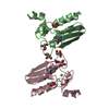
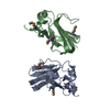
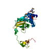
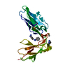
- PDB-3cq1: Structure of the DTDP-4-Keto-L-Rhamnose Reductase related protein... -
+ Open data

- Basic information
| Entry | Database: PDB / ID: 3cq1 | ||||||
|---|---|---|---|---|---|---|---|
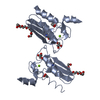
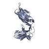
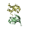
| Title | Structure of the DTDP-4-Keto-L-Rhamnose Reductase related protein (TT1362) from Thermus Thermophilus HB8 | ||||||
 Components Components | Putative uncharacterized protein TTHB138 | ||||||
 Keywords Keywords | OXIDOREDUCTASE / Thermus Thermophilus / DTDP-4-Keto-L-Rhamnose Reductase / Plasmid / Structural Genomics / NPPSFA / National Project on Protein Structural and Functional Analyses / RIKEN Structural Genomics/Proteomics Initiative / RSGI | ||||||
| Function / homology | : / Fe-S cluster assembly (FSCA) / MIP18 family-like / Iron-sulfur cluster assembly protein / Fe-S cluster assembly domain superfamily / GMP Synthetase; Chain A, domain 3 / 2-Layer Sandwich / Alpha Beta / MIP18 family-like domain-containing protein Function and homology information Function and homology information | ||||||
| Biological species |   Thermus thermophilus (bacteria) Thermus thermophilus (bacteria) | ||||||
| Method |  X-RAY DIFFRACTION / SYNCHROTRON / MOLECULAR REPLACEMENT / Resolution: 1.9 Å X-RAY DIFFRACTION / SYNCHROTRON / MOLECULAR REPLACEMENT / Resolution: 1.9 Å | ||||||
 Authors Authors | Jeyakanthan, J. / Satoh, S. / Kitamura, Y. / Yokoyama, S. / Kuramitsu, S. / RIKEN Structural Genomics/Proteomics Initiative (RSGI) | ||||||
 Citation Citation | Journal: To be Published Title: Structure of the DTDP-4-Keto-L-Rhamnose Reductase related protein (TT1362) from Thermus Thermophilus HB8 Authors: Jeyakanthan, J. / Satoh, S. / Kitamura, Y. / Yokoyama, S. / Kuramitsu, S. | ||||||
| History |
|
- Structure visualization
| Structure viewer | Molecule: MolmilJmol/JSmol |
|---|
- Downloads & links
-Download
| PDBx/mmCIF format | 3cq1.cif.gz | 33.9 KB | Display | PDBx/mmCIF format |
|---|---|---|---|---|
| PDB format | pdb3cq1.ent.gz | 22.8 KB | Display | PDB format |
| PDBx/mmJSON format | 3cq1.json.gz | Tree view | PDBx/mmJSON format | |
| Others |  Other downloads Other downloads |
-Validation report
| Summary document | 3cq1_validation.pdf.gz | 423.8 KB | Display | wwPDB validaton report |
|---|---|---|---|---|
| Full document | 3cq1_full_validation.pdf.gz | 426.7 KB | Display | |
| Data in XML | 3cq1_validation.xml.gz | 7 KB | Display | |
| Data in CIF | 3cq1_validation.cif.gz | 8.9 KB | Display | |
| Arichive directory | https://data.pdbj.org/pub/pdb/validation_reports/cq/3cq1ftp://data.pdbj.org/pub/pdb/validation_reports/cq/3cq1 | HTTPS FTP |
-Related structure data
| Related structure data | 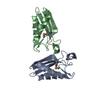 2cu6S S: Starting model for refinement |
|---|---|
| Similar structure data | |
| Other databases |


-Links
 PDBj
PDBj- Assembly
| Deposited unit | 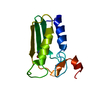
| ||||||||
|---|---|---|---|---|---|---|---|---|---|
| 1 | 
| ||||||||
| Unit cell |
| ||||||||
| Components on special symmetry positions |
|
-Components
| #1: Protein | Mass: 11495.020 Da / Num. of mol.: 1 Source method: isolated from a genetically manipulated source Source: (gene. exp.) Thermus thermophilus (bacteria) / Strain: HB8 / Plasmid: PET11A / Production host: |
|---|---|
| #2: Water | ChemComp-HOH /  Mass: 18.015 Da / Num. of mol.: 70 / Source method: isolated from a natural source / Formula: H2O Mass: 18.015 Da / Num. of mol.: 70 / Source method: isolated from a natural source / Formula: H2O |
| Has protein modification | Y |
-Experimental details
-Experiment
| Experiment | Method: X-RAY DIFFRACTION / Number of used crystals: 1 |
|---|
- Sample preparation
| Crystal | Density Matthews: 3.28 Å3/Da / Density % sol: 62.56 % |
|---|---|
| Crystal grow | Temperature: 293 K / Method: vapor diffusion, sitting drop / pH: 4 Details: 0.1M Acetate, 12% PEG400, 0.1M Mg Chloride, pH4.0, VAPOR DIFFUSION, SITTING DROP, temperature 293K |
-Data collection
| Diffraction | Mean temperature: 100 K |
|---|---|
| Diffraction source | Source: SYNCHROTRON / Site: SPring-8  / Beamline: BL26B2 / Wavelength: 1 Å / Beamline: BL26B2 / Wavelength: 1 Å |
| Detector | Type: RIGAKU / Detector: IMAGE PLATE / Date: Nov 8, 2004 / Details: RH Coated Bent-Cyrindrical MIRROR |
| Radiation | Monochromator: SI 1 1 1 DOUBLE CRYSTAL MONOCHROMATOR / Protocol: SINGLE WAVELENGTH / Monochromatic (M) / Laue (L): M / Scattering type: x-ray |
| Radiation wavelength | Wavelength: 1 Å / Relative weight: 1 |
| Reflection | Resolution: 1.9→50 Å / Num. obs: 12570 / % possible obs: 100 % / Biso Wilson estimate: 22.5 Å2 / Rmerge(I) obs: 0.07 / Rsym value: 0.08 |
| Reflection shell | Resolution: 1.9→1.97 Å / Rmerge(I) obs: 0.344 / Num. unique all: 1246 / Rsym value: 0.361 / % possible all: 100 |
- Processing
| Software |
| ||||||||||||||||||||
|---|---|---|---|---|---|---|---|---|---|---|---|---|---|---|---|---|---|---|---|---|---|
| Refinement | Method to determine structure: MOLECULAR REPLACEMENT Starting model: PDB ENTRY 2CU6 Resolution: 1.9→49.78 Å / Rfactor Rfree error: 0.009 / Data cutoff high absF: 1574972.35 / Data cutoff low absF: 0 / Isotropic thermal model: RESTRAINED / Cross valid method: THROUGHOUT / σ(F): 0 / Stereochemistry target values: Engh & Huber
| ||||||||||||||||||||
| Solvent computation | Solvent model: FLAT MODEL / Bsol: 58.9839 Å2 / ksol: 0.376525 e/Å3 | ||||||||||||||||||||
| Displacement parameters | Biso mean: 34.1 Å2
| ||||||||||||||||||||
| Refine analyze |
| ||||||||||||||||||||
| Refinement step | Cycle: LAST / Resolution: 1.9→49.78 Å
| ||||||||||||||||||||
| Refine LS restraints |
| ||||||||||||||||||||
| LS refinement shell | Resolution: 1.9→1.99 Å / Rfactor Rfree error: 0.026 / Total num. of bins used: 8
| ||||||||||||||||||||
| Xplor file |
|
