- PDB-3ch5: The crystal structure of the RanGDP-Nup153ZnF2 complex -
+
Open data
ID or keywords:
Loading...
-
Basic information
Entry
Database: PDB / ID: 3ch5
Title
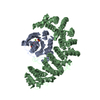
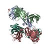
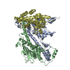
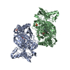
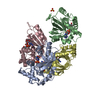
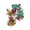
The crystal structure of the RanGDP-Nup153ZnF2 complex
Components
Fragment of Nuclear pore complex protein Nup153
GTP-binding nuclear protein Ran
Keywords
TRANSPORT PROTEIN / RanBP2 type C2-C2 Zinc Finger
Function / homology
Function and homology information
nucleoplasmic side of nuclear pore / Transcriptional regulation by small RNAs / Transport of the SLBP independent Mature mRNA / Transport of the SLBP Dependant Mature mRNA / Transport of Mature mRNA Derived from an Intronless Transcript / Transport of Mature mRNA derived from an Intron-Containing Transcript / snRNP Assembly / SUMOylation of ubiquitinylation proteins / Nuclear Pore Complex (NPC) Disassembly / SUMOylation of SUMOylation proteins ...nucleoplasmic side of nuclear pore / Transcriptional regulation by small RNAs / Transport of the SLBP independent Mature mRNA / Transport of the SLBP Dependant Mature mRNA / Transport of Mature mRNA Derived from an Intronless Transcript / Transport of Mature mRNA derived from an Intron-Containing Transcript / snRNP Assembly / SUMOylation of ubiquitinylation proteins / Nuclear Pore Complex (NPC) Disassembly / SUMOylation of SUMOylation proteins / SUMOylation of chromatin organization proteins / SUMOylation of RNA binding proteins / SUMOylation of DNA replication proteins / Regulation of Glucokinase by Glucokinase Regulatory Protein / SUMOylation of DNA damage response and repair proteins / negative regulation of RNA export from nucleus / Regulation of HSF1-mediated heat shock response / annulate lamellae / nuclear pore complex assembly / RNA nuclear export complex / pre-miRNA export from nucleus / snRNA import into nucleus / cellular response to mineralocorticoid stimulus / manchette / nuclear inclusion body / nuclear pore nuclear basket / Regulation of cholesterol biosynthesis by SREBP (SREBF) / importin-alpha family protein binding / structural constituent of nuclear pore / Rev-mediated nuclear export of HIV RNA / Nuclear import of Rev protein / protein localization to nucleolus / NEP/NS2 Interacts with the Cellular Export Machinery / RNA export from nucleus / tRNA processing in the nucleus / GTP metabolic process / Postmitotic nuclear pore complex (NPC) reformation / protein-containing complex localization / nuclear localization sequence binding / MicroRNA (miRNA) biogenesis / DNA metabolic process / dynein intermediate chain binding / mitotic sister chromatid segregation / viral process / spermatid development / ribosomal large subunit export from nucleus / nuclear pore / mRNA transport / sperm flagellum / ribosomal subunit export from nucleus / protein-membrane adaptor activity / ribosomal small subunit export from nucleus / protein export from nucleus / nuclear periphery / mitotic spindle organization / male germ cell nucleus / hippocampus development / centriole / molecular condensate scaffold activity / Transcriptional regulation by small RNAs / positive regulation of protein import into nucleus / recycling endosome / small GTPase binding / protein import into nucleus / melanosome / GDP binding / nuclear envelope / mitotic cell cycle / actin cytoskeleton organization / double-stranded DNA binding / G protein activity / midbody / nuclear membrane / amyloid fibril formation / Hydrolases; Acting on acid anhydrides; Acting on GTP to facilitate cellular and subcellular movement / cadherin binding / protein heterodimerization activity / protein domain specific binding / cell division / GTPase activity / chromatin binding / chromatin / GTP binding / protein-containing complex binding / nucleolus / magnesium ion binding / protein-containing complex / RNA binding / extracellular exosome / zinc ion binding / nucleoplasm / membrane / identical protein binding / nucleus / cytoplasm / cytosol Similarity search - Function
Nucleoporin Nup153, N-terminal / Retro-transposon transporting motif / Nucleoporin Nup153-like / Retro-transposon transporting motif / Nuclear pore complex protein / Ran GTPase / Small GTPase Ran-type domain profile. / Zinc finger domain / Zn-finger in Ran binding protein and others / Zinc finger RanBP2 type profile. ...Nucleoporin Nup153, N-terminal / Retro-transposon transporting motif / Nucleoporin Nup153-like / Retro-transposon transporting motif / Nuclear pore complex protein / Ran GTPase / Small GTPase Ran-type domain profile. / Zinc finger domain / Zn-finger in Ran binding protein and others / Zinc finger RanBP2 type profile. / Zinc finger, RanBP2-type superfamily / Zinc finger RanBP2-type signature. / Zinc finger, RanBP2-type / Ran (Ras-related nuclear proteins) /TC4 subfamily of small GTPases / Rho (Ras homology) subfamily of Ras-like small GTPases / Ras subfamily of RAS small GTPases / Small GTPase / Ras family / Rab subfamily of small GTPases / Small GTP-binding protein domain / P-loop containing nucleotide triphosphate hydrolases / Rossmann fold / P-loop containing nucleoside triphosphate hydrolase / 3-Layer(aba) Sandwich / Alpha Beta Similarity search - Domain/homology
GUANOSINE-5'-DIPHOSPHATE / Nuclear pore complex protein Nup153 / GTP-binding nuclear protein Ran Similarity search - Component
Biological species
Homo sapiens (human) Rattus norvegicus (Norway rat)
Mass: 18.015 Da / Num. of mol.: 71 / Source method: isolated from a natural source / Formula: H2O
-
Experimental details
-
Experiment
Experiment
Method: X-RAY DIFFRACTION / Number of used crystals: 1
-
Sample preparation
Crystal
Density Matthews: 2.71 Å3/Da / Density % sol: 54.57 %
Crystal grow
Temperature: 293 K / Method: vapor diffusion, hanging drop / pH: 5.5 Details: 2 M ammonium sulfate, 0.1 M Bis Tris, pH 5.5, VAPOR DIFFUSION, HANGING DROP, temperature 293K
In the structure databanks used in Yorodumi, some data are registered as the other names, "COVID-19 virus" and "2019-nCoV". Here are the details of the virus and the list of structure data.
Jan 31, 2019. EMDB accession codes are about to change! (news from PDBe EMDB page)
EMDB accession codes are about to change! (news from PDBe EMDB page)
The allocation of 4 digits for EMDB accession codes will soon come to an end. Whilst these codes will remain in use, new EMDB accession codes will include an additional digit and will expand incrementally as the available range of codes is exhausted. The current 4-digit format prefixed with “EMD-” (i.e. EMD-XXXX) will advance to a 5-digit format (i.e. EMD-XXXXX), and so on. It is currently estimated that the 4-digit codes will be depleted around Spring 2019, at which point the 5-digit format will come into force.
The EM Navigator/Yorodumi systems omit the EMD- prefix.
Related info.:Q: What is EMD? / ID/Accession-code notation in Yorodumi/EM Navigator
Yorodumi is a browser for structure data from EMDB, PDB, SASBDB, etc.
This page is also the successor to EM Navigator detail page, and also detail information page/front-end page for Omokage search.
The word "yorodu" (or yorozu) is an old Japanese word meaning "ten thousand". "mi" (miru) is to see.
Related info.:EMDB / PDB / SASBDB / Comparison of 3 databanks / Yorodumi Search / Aug 31, 2016. New EM Navigator & Yorodumi / Yorodumi Papers / Jmol/JSmol / Function and homology information / Changes in new EM Navigator and Yorodumi
 Movie
Movie Controller


 Open data
 Basic information
 Components
 Keywords
 Function and homology information
 Homo sapiens (human)
 X-RAY DIFFRACTION /
  Authors
 Citation
 Structure visualization
 Downloads & links
 Other downloads
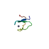


 PDBj
PDBj


 Assembly


 Mass: 24.305 Da / Num. of mol.: 1 / Source method: obtained synthetically / Formula: Mg
 Mass: 96.063 Da / Num. of mol.: 1 / Source method: obtained synthetically / Formula: SO4
 Type: RNA linking / Mass: 443.201 Da / Num. of mol.: 1 / Source method: obtained synthetically / Formula: C10H15N5O11P2 / Comment: GDP, energy-carrying molecule*YM
 Mass: 65.409 Da / Num. of mol.: 1 / Source method: obtained synthetically / Formula: Zn
 Sample preparation
 / Beamline: X10SA / Wavelength: 1.2726 Å
 Processing