Regulation of PD-L1(CD274) transcription / cellular heat acclimation / NURF complex / NuRD complex / regulation of cell fate specification / negative regulation of stem cell population maintenance / ESC/E(Z) complex / regulation of stem cell differentiation / response to steroid hormone / histone deacetylase complex ...Regulation of PD-L1(CD274) transcription / cellular heat acclimation / NURF complex / NuRD complex / regulation of cell fate specification / negative regulation of stem cell population maintenance / ESC/E(Z) complex / regulation of stem cell differentiation / response to steroid hormone / histone deacetylase complex / positive regulation of stem cell population maintenance / Transcriptional Regulation by E2F6 / RNA Polymerase I Transcription Initiation / Transcriptional regulation of brown and beige adipocyte differentiation by EBF2 / Regulation of TP53 Activity through Acetylation / negative regulation of megakaryocyte differentiation / protein localization to CENP-A containing chromatin / Replacement of protamines by nucleosomes in the male pronucleus / CENP-A containing nucleosome / Packaging Of Telomere Ends / Recognition and association of DNA glycosylase with site containing an affected purine / Cleavage of the damaged purine / Deposition of new CENPA-containing nucleosomes at the centromere / telomere organization / Recognition and association of DNA glycosylase with site containing an affected pyrimidine / Cleavage of the damaged pyrimidine / RNA Polymerase I Promoter Opening / Inhibition of DNA recombination at telomere / negative regulation of cell migration / Assembly of the ORC complex at the origin of replication / Meiotic synapsis / SUMOylation of chromatin organization proteins / Regulation of endogenous retroelements by the Human Silencing Hub (HUSH) complex / Regulation of PTEN gene transcription / DNA methylation / Condensation of Prophase Chromosomes / Chromatin modifications during the maternal to zygotic transition (MZT) / SIRT1 negatively regulates rRNA expression / HCMV Late Events / ERCC6 (CSB) and EHMT2 (G9a) positively regulate rRNA expression / PRC2 methylates histones and DNA / Regulation of endogenous retroelements by KRAB-ZFP proteins / Defective pyroptosis / HDACs deacetylate histones / Regulation of endogenous retroelements by Piwi-interacting RNAs (piRNAs) / RNA Polymerase I Promoter Escape / negative regulation of transforming growth factor beta receptor signaling pathway / Nonhomologous End-Joining (NHEJ) / Transcriptional regulation by small RNAs / HDMs demethylate histones / Formation of the beta-catenin:TCF transactivating complex / Activated PKN1 stimulates transcription of AR (androgen receptor) regulated genes KLK2 and KLK3 / RUNX1 regulates genes involved in megakaryocyte differentiation and platelet function / negative regulation of cell growth / brain development / NoRC negatively regulates rRNA expression / Negative Regulation of CDH1 Gene Transcription / G2/M DNA damage checkpoint / PKMTs methylate histone lysines / B-WICH complex positively regulates rRNA expression / DNA Damage/Telomere Stress Induced Senescence / Meiotic recombination / Pre-NOTCH Transcription and Translation / Activation of anterior HOX genes in hindbrain development during early embryogenesis / Transcriptional regulation of granulopoiesis / RMTs methylate histone arginines / HCMV Early Events / structural constituent of chromatin / nucleosome / nucleosome assembly / heterochromatin formation / HATs acetylate histones / Recruitment and ATM-mediated phosphorylation of repair and signaling proteins at DNA double strand breaks / MLL4 and MLL3 complexes regulate expression of PPARG target genes in adipogenesis and hepatic steatosis / chromatin organization / RUNX1 regulates transcription of genes involved in differentiation of HSCs / Neddylation / Processing of DNA double-strand break ends / Senescence-Associated Secretory Phenotype (SASP) / histone binding / Oxidative Stress Induced Senescence / Estrogen-dependent gene expression / Potential therapeutics for SARS / DNA replication / chromosome, telomeric region / chromatin remodeling / Amyloid fiber formation / protein heterodimerization activity / negative regulation of DNA-templated transcription / regulation of DNA-templated transcription / positive regulation of DNA-templated transcription / DNA-templated transcription / negative regulation of transcription by RNA polymerase II / protein-containing complex / DNA binding / RNA binding / extracellular exosome / extracellular region / nucleoplasm / membrane Similarity search - Function
Histone-binding protein RBBP4, N-terminal / Histone-binding protein RBBP4 or subunit C of CAF1 complex / : / YVTN repeat-like/Quinoprotein amine dehydrogenase / 7 Propeller / Methylamine Dehydrogenase; Chain H / TATA box binding protein associated factor / TATA box binding protein associated factor (TAF), histone-like fold domain / Histone H4, conserved site / Histone H4 signature. ...Histone-binding protein RBBP4, N-terminal / Histone-binding protein RBBP4 or subunit C of CAF1 complex / : / YVTN repeat-like/Quinoprotein amine dehydrogenase / 7 Propeller / Methylamine Dehydrogenase; Chain H / TATA box binding protein associated factor / TATA box binding protein associated factor (TAF), histone-like fold domain / Histone H4, conserved site / Histone H4 signature. / Histone H4 / Histone H4 / CENP-T/Histone H4, histone fold / Centromere kinetochore component CENP-T histone fold / Histone-fold / WD domain, G-beta repeat / G-protein beta WD-40 repeat / WD40 repeat, conserved site / Trp-Asp (WD) repeats signature. / Trp-Asp (WD) repeats profile. / Trp-Asp (WD) repeats circular profile. / WD40 repeats / WD40 repeat / WD40-repeat-containing domain superfamily / WD40/YVTN repeat-like-containing domain superfamily / Mainly Beta Similarity search - Domain/homology
Resolution: 2.4→2.49 Å / Redundancy: 3.6 % / Rmerge(I) obs: 0.468 / Mean I/σ(I) obs: 1.9 / Rsym value: 0.409 / % possible all: 98
-
Processing
Software
Name
Version
Classification
REFMAC
5.2.0019
refinement
ADSC
Quantum
datacollection
DENZO
datareduction
HKL-2000
datascaling
PHASES
phasing
Refinement
Method to determine structure: MOLECULAR REPLACEMENT Starting model: SAD data phased model Resolution: 2.4→19.53 Å / Cor.coef. Fo:Fc: 0.942 / Cor.coef. Fo:Fc free: 0.909 / SU B: 4.405 / SU ML: 0.108 / Isotropic thermal model: Isotropic / Cross valid method: THROUGHOUT / ESU R: 0.426 / ESU R Free: 0.269 / Stereochemistry target values: MAXIMUM LIKELIHOOD / Details: HYDROGENS HAVE BEEN ADDED IN THE RIDING POSITIONS
Rfactor
Num. reflection
% reflection
Selection details
Rfree
0.24862
935
5.1 %
RANDOM
Rwork
0.18418
-
-
-
obs
0.18745
17375
99.95 %
-
all
-
17375
-
-
Solvent computation
Ion probe radii: 0.8 Å / Shrinkage radii: 0.8 Å / VDW probe radii: 1.2 Å / Solvent model: BABINET MODEL WITH MASK
Displacement parameters
Biso mean: 31.2 Å2
Baniso -1
Baniso -2
Baniso -3
1-
-1 Å2
0 Å2
0 Å2
2-
-
1.2 Å2
0 Å2
3-
-
-
-0.19 Å2
Refine analyze
Free
Obs
Luzzati coordinate error
0.268 Å
0.094 Å
Luzzati d res low
-
6 Å
Luzzati sigma a
0.229 Å
0.251 Å
Refinement step
Cycle: LAST / Resolution: 2.4→19.53 Å
Protein
Nucleic acid
Ligand
Solvent
Total
Num. atoms
3186
0
1
210
3397
Refine LS restraints
Refine-ID
Type
Dev ideal
Dev ideal target
Number
X-RAY DIFFRACTION
r_bond_refined_d
0.003
0.021
3273
X-RAY DIFFRACTION
r_bond_other_d
X-RAY DIFFRACTION
r_angle_refined_deg
0.75
1.926
4458
X-RAY DIFFRACTION
r_angle_other_deg
X-RAY DIFFRACTION
r_dihedral_angle_1_deg
5.663
5
395
X-RAY DIFFRACTION
r_dihedral_angle_2_deg
32.463
24.845
161
X-RAY DIFFRACTION
r_dihedral_angle_3_deg
16.715
15
538
X-RAY DIFFRACTION
r_dihedral_angle_4_deg
14.773
15
14
X-RAY DIFFRACTION
r_chiral_restr
0.061
0.2
485
X-RAY DIFFRACTION
r_gen_planes_refined
0.002
0.02
2515
X-RAY DIFFRACTION
r_gen_planes_other
X-RAY DIFFRACTION
r_nbd_refined
0.213
0.3
1397
X-RAY DIFFRACTION
r_nbd_other
X-RAY DIFFRACTION
r_nbtor_refined
0.319
0.5
2143
X-RAY DIFFRACTION
r_nbtor_other
X-RAY DIFFRACTION
r_xyhbond_nbd_refined
0.174
0.5
346
X-RAY DIFFRACTION
r_xyhbond_nbd_other
X-RAY DIFFRACTION
r_metal_ion_refined
X-RAY DIFFRACTION
r_metal_ion_other
X-RAY DIFFRACTION
r_symmetry_vdw_refined
0.127
0.3
31
X-RAY DIFFRACTION
r_symmetry_vdw_other
X-RAY DIFFRACTION
r_symmetry_hbond_refined
0.187
0.5
12
X-RAY DIFFRACTION
r_symmetry_hbond_other
X-RAY DIFFRACTION
r_symmetry_metal_ion_refined
X-RAY DIFFRACTION
r_symmetry_metal_ion_other
X-RAY DIFFRACTION
r_mcbond_it
4.836
2
2030
X-RAY DIFFRACTION
r_mcbond_other
X-RAY DIFFRACTION
r_mcangle_it
6.542
3
3223
X-RAY DIFFRACTION
r_scbond_it
5.491
2
1421
X-RAY DIFFRACTION
r_scangle_it
7.295
3
1235
X-RAY DIFFRACTION
r_rigid_bond_restr
X-RAY DIFFRACTION
r_sphericity_free
X-RAY DIFFRACTION
r_sphericity_bonded
LS refinement shell
Resolution: 2.4→2.461 Å / Total num. of bins used: 20
Rfactor
Num. reflection
% reflection
Rfree
0.279
69
-
Rwork
0.192
1251
-
obs
-
-
99.7 %
+
About Yorodumi
-
News
-
Feb 9, 2022. New format data for meta-information of EMDB entries
New format data for meta-information of EMDB entries
Version 3 of the EMDB header file is now the official format.
The previous official version 1.9 will be removed from the archive.
In the structure databanks used in Yorodumi, some data are registered as the other names, "COVID-19 virus" and "2019-nCoV". Here are the details of the virus and the list of structure data.
Jan 31, 2019. EMDB accession codes are about to change! (news from PDBe EMDB page)
EMDB accession codes are about to change! (news from PDBe EMDB page)
The allocation of 4 digits for EMDB accession codes will soon come to an end. Whilst these codes will remain in use, new EMDB accession codes will include an additional digit and will expand incrementally as the available range of codes is exhausted. The current 4-digit format prefixed with “EMD-” (i.e. EMD-XXXX) will advance to a 5-digit format (i.e. EMD-XXXXX), and so on. It is currently estimated that the 4-digit codes will be depleted around Spring 2019, at which point the 5-digit format will come into force.
The EM Navigator/Yorodumi systems omit the EMD- prefix.
Related info.:Q: What is EMD? / ID/Accession-code notation in Yorodumi/EM Navigator
Yorodumi is a browser for structure data from EMDB, PDB, SASBDB, etc.
This page is also the successor to EM Navigator detail page, and also detail information page/front-end page for Omokage search.
The word "yorodu" (or yorozu) is an old Japanese word meaning "ten thousand". "mi" (miru) is to see.
Related info.:EMDB / PDB / SASBDB / Comparison of 3 databanks / Yorodumi Search / Aug 31, 2016. New EM Navigator & Yorodumi / Yorodumi Papers / Jmol/JSmol / Function and homology information / Changes in new EM Navigator and Yorodumi
 Movie
Movie Controller

 Yorodumi
 Open data
 Basic information
 Components
 Keywords
 Function and homology information
 Homo sapiens (human)
 X-RAY DIFFRACTION /
  Authors
 Citation
 Structure visualization
 Downloads & links
 Other downloads

 PDBj
PDBj


 Assembly

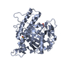
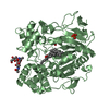
 Spodoptera frugiperda (fall armyworm) / References: UniProt: Q16576

 Mass: 74.922 Da / Num. of mol.: 1 / Source method: obtained synthetically / Formula: As
 Mass: 18.015 Da / Num. of mol.: 210 / Source method: isolated from a natural source / Formula: H2O
 Sample preparation
 / Beamline: ID29 / Wavelength: 0.9792 Å
 Processing