 Movie
Movie Controller

[English] 日本語
 Yorodumi
- PDB-3c0t: Structure of the Schizosaccharomyces pombe Mediator subcomplex Me... -
+ Open data

- Basic information
| Entry | Database: PDB / ID: 3c0t | ||||||
|---|---|---|---|---|---|---|---|
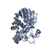
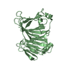
| Title | Structure of the Schizosaccharomyces pombe Mediator subcomplex Med8C/18 | ||||||
 Components Components |
| ||||||
 Keywords Keywords | TRANSCRIPTION / BETA BARREL / CHANNEL / PROTEIN-PROTEIN COMPLEX / Activator / Nucleus / Transcription regulation | ||||||
| Function / homology |  Function and homology information Function and homology informationcore mediator complex / mediator complex / positive regulation of transcription initiation by RNA polymerase II / transcription coregulator activity / transcription initiation at RNA polymerase II promoter / transcription coactivator activity / RNA polymerase II cis-regulatory region sequence-specific DNA binding / regulation of transcription by RNA polymerase II / nucleus Similarity search - Function | ||||||
| Biological species |  | ||||||
| Method |  X-RAY DIFFRACTION / SYNCHROTRON / MOLECULAR REPLACEMENT / Resolution: 2.4 Å X-RAY DIFFRACTION / SYNCHROTRON / MOLECULAR REPLACEMENT / Resolution: 2.4 Å | ||||||
 Authors Authors | Lariviere, L. / Seizl, M. / van Wageningen, S. / Roether, S. / van de Pasch, L. / Feldmann, H. / Straesser, K. / Hahn, S. / Holstege, C.P. / Cramer, P. | ||||||
 Citation Citation | Journal: Genes Dev. / Year: 2008 Title: Structure-system correlation identifies a gene regulatory Mediator submodule Authors: Lariviere, L. / Seizl, M. / van Wageningen, S. / Rother, S. / van de Pasch, L. / Feldmann, H. / Strasser, K. / Hahn, S. / Holstege, F.C.P. / Cramer, P. | ||||||
| History |
|
- Structure visualization
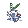
| Structure viewer | Molecule: MolmilJmol/JSmol |
|---|
- Downloads & links
-Download
| PDBx/mmCIF format | 3c0t.cif.gz | 58.5 KB | Display | PDBx/mmCIF format |
|---|---|---|---|---|
| PDB format | pdb3c0t.ent.gz | 43.2 KB | Display | PDB format |
| PDBx/mmJSON format | 3c0t.json.gz | Tree view | PDBx/mmJSON format | |
| Others |  Other downloads Other downloads |
-Validation report
| Arichive directory | https://data.pdbj.org/pub/pdb/validation_reports/c0/3c0tftp://data.pdbj.org/pub/pdb/validation_reports/c0/3c0t | HTTPS FTP |
|---|
-Related structure data
| Related structure data | |
|---|---|
| Similar structure data |


-Links
 PDBj
PDBj- Assembly
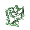
| Deposited unit | 
| ||||||||
|---|---|---|---|---|---|---|---|---|---|
| 1 |
| ||||||||
| Unit cell |
|
-Components
| #1: Protein | Mass: 24056.377 Da / Num. of mol.: 1 Source method: isolated from a genetically manipulated source Source: (gene. exp.) Gene: med18 / Production host:  |
|---|---|
| #2: Protein/peptide | Mass: 3859.357 Da / Num. of mol.: 1 / Fragment: C-terminal domain, UNP Residues 180-200 Source method: isolated from a genetically manipulated source Source: (gene. exp.) Gene: med8 / Production host: |
| #3: Water | ChemComp-HOH /  Mass: 18.015 Da / Num. of mol.: 62 / Source method: isolated from a natural source / Formula: H2O Mass: 18.015 Da / Num. of mol.: 62 / Source method: isolated from a natural source / Formula: H2O |
-Experimental details
-Experiment
| Experiment | Method: X-RAY DIFFRACTION / Number of used crystals: 1 |
|---|
- Sample preparation
| Crystal | Density Matthews: 4.37 Å3/Da / Density % sol: 71.88 % |
|---|---|
| Crystal grow | Temperature: 293 K / Method: vapor diffusion, hanging drop / pH: 8.5 Details: 100mM Tris pH 8.5, 2M sodium formate, VAPOR DIFFUSION, HANGING DROP, temperature 293K |
-Data collection
| Diffraction | Mean temperature: 100 K |
|---|---|
| Diffraction source | Source: SYNCHROTRON / Site: SLS  / Beamline: X06SA / Wavelength: 0.979 Å / Beamline: X06SA / Wavelength: 0.979 Å |
| Detector | Type: MARMOSAIC 225 mm CCD / Detector: CCD / Date: Oct 1, 2006 |
| Radiation | Monochromator: SI 111 / Protocol: SINGLE WAVELENGTH / Monochromatic (M) / Laue (L): M / Scattering type: x-ray |
| Radiation wavelength | Wavelength: 0.979 Å / Relative weight: 1 |
| Reflection | Resolution: 2.4→20 Å / Num. all: 19282 / Num. obs: 19282 / % possible obs: 99.4 % / Observed criterion σ(F): 0 / Observed criterion σ(I): 0 / Redundancy: 11 % / Rsym value: 0.084 / Net I/σ(I): 21.4 |
| Reflection shell | Resolution: 2.4→2.53 Å / Redundancy: 11.2 % / Mean I/σ(I) obs: 4.8 / Num. unique all: 2806 / Rsym value: 0.379 / % possible all: 99.4 |
- Processing
| Software |
| ||||||||||||||||||||
|---|---|---|---|---|---|---|---|---|---|---|---|---|---|---|---|---|---|---|---|---|---|
| Refinement | Method to determine structure: MOLECULAR REPLACEMENT Starting model: The starting model is the structure of Med18 alone, which has not been deposited. Resolution: 2.4→20 Å / Isotropic thermal model: isotropic / σ(F): 0 / Stereochemistry target values: Engh & Huber
| ||||||||||||||||||||
| Refinement step | Cycle: LAST / Resolution: 2.4→20 Å
| ||||||||||||||||||||
| Refine LS restraints |
|
