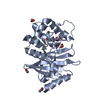
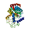
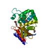
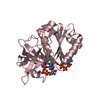
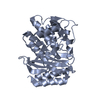
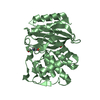
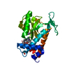
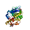
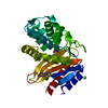
- PDB-3bxd: Crystal structure of Mouse Myo-inositol oxygenase (re-refined) -
+
Open data
ID or keywords:
Loading...
-
Basic information
Entry
Database: PDB / ID: 3bxd
Title
Crystal structure of Mouse Myo-inositol oxygenase (re-refined)
Components
INOSITOL OXYGENASE
Keywords
OXIDOREDUCTASE / PROTEIN-SUBSTRATE COMPLEX / HD DOMAIN FOLD / DIIRON
Function / homology
Function and homology information
inositol oxygenase / inositol oxygenase activity / Synthesis of IP2, IP, and Ins in the cytosol / inositol catabolic process / alcohol dehydrogenase (NADP+) activity / oxidoreductase activity, acting on NAD(P)H / inclusion body / ferric iron binding / NADP binding / oxidoreductase activity / cytoplasm Similarity search - Function
Inositol oxygenase / Myo-inositol oxygenase / Aldo/keto reductase family putative active site signature. / Aldo/keto reductase, conserved site Similarity search - Domain/homology
THIS ENTRY 3BXD REFLECTS AN ALTERNATIVE MODELING OF THE STRUCTURAL DATA IN R2HUOSF. IN PDB ENTRY ... THIS ENTRY 3BXD REFLECTS AN ALTERNATIVE MODELING OF THE STRUCTURAL DATA IN R2HUOSF. IN PDB ENTRY 3BXD INFORMATION IN REMARK 200 SERIES IS BASED ON THE EXPERIMENT DESCRIBED IN PDB ENTRY 2HUO. ORIGINAL DATA DETERMINED BY AUTHOR J.WANG
In the structure databanks used in Yorodumi, some data are registered as the other names, "COVID-19 virus" and "2019-nCoV". Here are the details of the virus and the list of structure data.
Jan 31, 2019. EMDB accession codes are about to change! (news from PDBe EMDB page)
EMDB accession codes are about to change! (news from PDBe EMDB page)
The allocation of 4 digits for EMDB accession codes will soon come to an end. Whilst these codes will remain in use, new EMDB accession codes will include an additional digit and will expand incrementally as the available range of codes is exhausted. The current 4-digit format prefixed with “EMD-” (i.e. EMD-XXXX) will advance to a 5-digit format (i.e. EMD-XXXXX), and so on. It is currently estimated that the 4-digit codes will be depleted around Spring 2019, at which point the 5-digit format will come into force.
The EM Navigator/Yorodumi systems omit the EMD- prefix.
Related info.:Q: What is EMD? / ID/Accession-code notation in Yorodumi/EM Navigator
Yorodumi is a browser for structure data from EMDB, PDB, SASBDB, etc.
This page is also the successor to EM Navigator detail page, and also detail information page/front-end page for Omokage search.
The word "yorodu" (or yorozu) is an old Japanese word meaning "ten thousand". "mi" (miru) is to see.
Related info.:EMDB / PDB / SASBDB / Comparison of 3 databanks / Yorodumi Search / Aug 31, 2016. New EM Navigator & Yorodumi / Yorodumi Papers / Jmol/JSmol / Function and homology information / Changes in new EM Navigator and Yorodumi
 Movie
Movie Controller


 Open data
 Basic information
 Components
 Keywords
 Function and homology information
 X-RAY DIFFRACTION / Resolution: 2 Å
  Authors
 Citation
 Structure visualization
 Downloads & links
 Other downloads
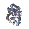


 PDBj
PDBj


 Assembly
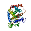


 Mass: 55.845 Da / Num. of mol.: 2 / Source method: obtained synthetically / Formula: Fe
 Mass: 17.007 Da / Num. of mol.: 1 / Source method: obtained synthetically / Formula: HO
 Mass: 180.156 Da / Num. of mol.: 1 / Source method: obtained synthetically / Formula: C6H12O6
 Mass: 46.025 Da / Num. of mol.: 1 / Source method: obtained synthetically / Formula: CH2O2
 Sample preparation
 Processing