 Movie
Movie Controller

[English] 日本語
 Yorodumi
- PDB-4m5r: High-resolution influenza 2009 H1N1 endonuclease bound to 4-(1H-I... -
+ Open data

- Basic information
| Entry | Database: PDB / ID: 4m5r | ||||||
|---|---|---|---|---|---|---|---|
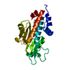
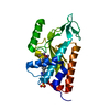
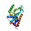
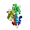
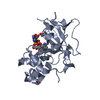
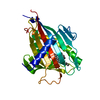
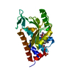
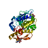
| Title | High-resolution influenza 2009 H1N1 endonuclease bound to 4-(1H-IMIDAZOL-1-YL)PHENOL | ||||||
 Components Components | Polymerase PA | ||||||
 Keywords Keywords | RNA BINDING PROTEIN/INHIBITOR / CAP-SNATCHING / RNA BINDING PROTEIN-INHIBITOR complex | ||||||
| Function / homology | : / 4-(1H-IMIDAZOL-1-YL)PHENOL / :  Function and homology information Function and homology information | ||||||
| Biological species |   Influenza A virus Influenza A virus | ||||||
| Method |  X-RAY DIFFRACTION / SYNCHROTRON / MOLECULAR REPLACEMENT / Resolution: 1.4 Å X-RAY DIFFRACTION / SYNCHROTRON / MOLECULAR REPLACEMENT / Resolution: 1.4 Å | ||||||
 Authors Authors | Bauman, J.D. / Patel, D. / Das, K. / Arnold, E. | ||||||
 Citation Citation | Journal: Acs Chem.Biol. / Year: 2013 Title: Crystallographic fragment screening and structure-based optimization yields a new class of influenza endonuclease inhibitors. Authors: Bauman, J.D. / Patel, D. / Baker, S.F. / Vijayan, R.S. / Xiang, A. / Parhi, A.K. / Martinez-Sobrido, L. / Lavoie, E.J. / Das, K. / Arnold, E. | ||||||
| History |
|
- Structure visualization
| Structure viewer | Molecule: MolmilJmol/JSmol |
|---|
- Downloads & links
-Download
| PDBx/mmCIF format | 4m5r.cif.gz | 143.9 KB | Display | PDBx/mmCIF format |
|---|---|---|---|---|
| PDB format | pdb4m5r.ent.gz | 114.3 KB | Display | PDB format |
| PDBx/mmJSON format | 4m5r.json.gz | Tree view | PDBx/mmJSON format | |
| Others |  Other downloads Other downloads |
-Validation report
| Arichive directory | https://data.pdbj.org/pub/pdb/validation_reports/m5/4m5rftp://data.pdbj.org/pub/pdb/validation_reports/m5/4m5r | HTTPS FTP |
|---|
-Related structure data
| Related structure data |  4ln7C  4m4qC  4m5oC  4m5qC  4m5vC  4mk1C  4mk2C  4mk5C C: citing same article ( |
|---|---|
| Similar structure data |


-Links
 PDBj
PDBj- Assembly
| Deposited unit | 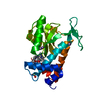
| ||||||||
|---|---|---|---|---|---|---|---|---|---|
| 1 |
| ||||||||
| 2 | 
| ||||||||
| Unit cell |
| ||||||||
| Components on special symmetry positions |
|
-Components
-Protein , 1 types, 1 molecules A
| #1: Protein | Mass: 28505.270 Da / Num. of mol.: 1 / Fragment: unp residues 1-204 Source method: isolated from a genetically manipulated source Source: (gene. exp.) Influenza A virus (A/Lima/WRAIR1695P/2009(H1N1))Gene: PA / Plasmid: PCDF-2 / Production host:  |
|---|
-Non-polymers , 5 types, 233 molecules 


| #2: Chemical | ChemComp-MN /  Mass: 54.938 Da / Num. of mol.: 4 / Source method: obtained synthetically / Formula: Mn Mass: 54.938 Da / Num. of mol.: 4 / Source method: obtained synthetically / Formula: Mn#3: Chemical |  Mass: 160.173 Da / Num. of mol.: 2 / Source method: obtained synthetically / Formula: C9H8N2O Mass: 160.173 Da / Num. of mol.: 2 / Source method: obtained synthetically / Formula: C9H8N2O#4: Chemical | ChemComp-SO4 / |  Mass: 96.063 Da / Num. of mol.: 1 / Source method: obtained synthetically / Formula: SO4 Mass: 96.063 Da / Num. of mol.: 1 / Source method: obtained synthetically / Formula: SO4#5: Chemical | ChemComp-EDO / |  Mass: 62.068 Da / Num. of mol.: 1 / Source method: obtained synthetically / Formula: C2H6O2 Mass: 62.068 Da / Num. of mol.: 1 / Source method: obtained synthetically / Formula: C2H6O2#6: Water | ChemComp-HOH / | Mass: 18.015 Da / Num. of mol.: 225 / Source method: isolated from a natural source / Formula: H2O |
|---|
-Experimental details
-Experiment
| Experiment | Method: X-RAY DIFFRACTION / Number of used crystals: 1 |
|---|
- Sample preparation
| Crystal | Density Matthews: 2.61 Å3/Da / Density % sol: 52.86 % |
|---|---|
| Crystal grow | Temperature: 293 K / Method: vapor diffusion, hanging drop / pH: 7.3 Details: 200 MM MES, 27% PEG8K, 200 MM AMMONIUM SULFATE, 1 MM MANGANESE CHLORIDE, 10 MM MAGNESIUM ACETATE, 10 MM TAURINE, AND 50 MM SODIUM FLUORIDE, pH 7.3, VAPOR DIFFUSION, HANGING DROP, temperature 293K |
-Data collection
| Diffraction | Mean temperature: 100 K |
|---|---|
| Diffraction source | Source: SYNCHROTRON / Site: CHESS  / Beamline: F1 / Wavelength: 0.917 / Wavelength: 0.917 Å / Beamline: F1 / Wavelength: 0.917 / Wavelength: 0.917 Å |
| Detector | Type: ADSC QUANTUM 270 / Detector: CCD / Date: Dec 2, 2010 |
| Radiation | Monochromator: HORIZONTAL BENT SI(111), ASYMMETRICALLY CUT WITH WATER COOLED CU BLOCK Protocol: SINGLE WAVELENGTH / Monochromatic (M) / Laue (L): M / Scattering type: x-ray |
| Radiation wavelength | Wavelength: 0.917 Å / Relative weight: 1 |
| Reflection | Resolution: 1.4→50 Å / Num. obs: 65059 / % possible obs: 99.1 % / Observed criterion σ(I): -3 / Redundancy: 6.6 % / Rmerge(I) obs: 0.084 / Net I/σ(I): 26.4 |
| Reflection shell | Resolution: 1.4→1.43 Å / Redundancy: 3.7 % / Rmerge(I) obs: 0.677 / Mean I/σ(I) obs: 1.66 / % possible all: 97.3 |
- Processing
| Software |
| |||||||||||||||||||||||||||||||||||||||||||||||||||||||||||||||
|---|---|---|---|---|---|---|---|---|---|---|---|---|---|---|---|---|---|---|---|---|---|---|---|---|---|---|---|---|---|---|---|---|---|---|---|---|---|---|---|---|---|---|---|---|---|---|---|---|---|---|---|---|---|---|---|---|---|---|---|---|---|---|---|---|
| Refinement | Method to determine structure: MOLECULAR REPLACEMENT / Resolution: 1.4→31.943 Å / SU ML: 0.19 / σ(F): 1.34 / Phase error: 22.77 / Stereochemistry target values: ML
| |||||||||||||||||||||||||||||||||||||||||||||||||||||||||||||||
| Solvent computation | Shrinkage radii: 0.9 Å / VDW probe radii: 1.11 Å / Solvent model: FLAT BULK SOLVENT MODEL | |||||||||||||||||||||||||||||||||||||||||||||||||||||||||||||||
| Refinement step | Cycle: LAST / Resolution: 1.4→31.943 Å
| |||||||||||||||||||||||||||||||||||||||||||||||||||||||||||||||
| Refine LS restraints |
| |||||||||||||||||||||||||||||||||||||||||||||||||||||||||||||||
| LS refinement shell |
|
