 Movie
Movie Controller

[English] 日本語
 Yorodumi
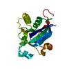
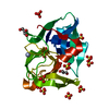
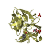
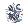
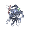
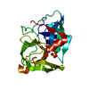
- PDB-3bqf: Structure of the central domain (MsrA) of Neisseria meningitidis ... -
+ Open data

- Basic information
| Entry | Database: PDB / ID: 3bqf | ||||||
|---|---|---|---|---|---|---|---|
| Title | Structure of the central domain (MsrA) of Neisseria meningitidis PilB (complex with a substrate) | ||||||
 Components Components | Peptide methionine sulfoxide reductase msrA/msrB | ||||||
 Keywords Keywords | OXIDOREDUCTASE / PILB / Methionine sulfoxide reductase A / complex with a substrate / Electron transport / Multifunctional enzyme / Redox-active center / Transport | ||||||
| Function / homology |  Function and homology information Function and homology informationpeptide-methionine (R)-S-oxide reductase / peptide-methionine (R)-S-oxide reductase activity / L-methionine (S)-S-oxide reductase activity / peptide-methionine (S)-S-oxide reductase / peptide-methionine (S)-S-oxide reductase activity / cellular response to oxidative stress / cytoplasm Similarity search - Function | ||||||
| Biological species |  Neisseria meningitidis (bacteria) Neisseria meningitidis (bacteria) | ||||||
| Method |  X-RAY DIFFRACTION / SYNCHROTRON / MOLECULAR REPLACEMENT / Resolution: 2.24 Å X-RAY DIFFRACTION / SYNCHROTRON / MOLECULAR REPLACEMENT / Resolution: 2.24 Å | ||||||
 Authors Authors | Ranaivoson, F.M. / Kauffmann, B. / Favier, F. | ||||||
 Citation Citation | Journal: J.Mol.Biol. / Year: 2008 Title: A structural analysis of the catalytic mechanism of methionine sulfoxide reductase A from Neisseria meningitidis Authors: Ranaivoson, F.M. / Antoine, M. / Kauffmann, B. / Boschi-Muller, S. / Aubry, A. / Branlant, G. / Favier, F. | ||||||
| History |
|
- Structure visualization
| Structure viewer | Molecule: MolmilJmol/JSmol |
|---|
- Downloads & links
-Download
| PDBx/mmCIF format | 3bqf.cif.gz | 50.1 KB | Display | PDBx/mmCIF format |
|---|---|---|---|---|
| PDB format | pdb3bqf.ent.gz | 33.8 KB | Display | PDB format |
| PDBx/mmJSON format | 3bqf.json.gz | Tree view | PDBx/mmJSON format | |
| Others |  Other downloads Other downloads |
-Validation report
| Arichive directory | https://data.pdbj.org/pub/pdb/validation_reports/bq/3bqfftp://data.pdbj.org/pub/pdb/validation_reports/bq/3bqf | HTTPS FTP |
|---|
-Related structure data
| Related structure data |  3bqeSC  3bqgC  3bqhC S: Starting model for refinement C: citing same article ( |
|---|---|
| Similar structure data |


-Links
 PDBj
PDBj- Assembly
| Deposited unit | 
| ||||||||
|---|---|---|---|---|---|---|---|---|---|
| 1 |
| ||||||||
| Unit cell |
|
-Components
| #1: Protein | Mass: 21908.264 Da / Num. of mol.: 1 Fragment: Msra Domain, Peptide methionine sulfoxide reductase A, UNP residues 196-389 Mutation: C207S Source method: isolated from a genetically manipulated source Source: (gene. exp.) Neisseria meningitidis (bacteria) / Strain: Z2491 / Gene: pilB / Plasmid: pSKPILBMsrA / Production host: References: UniProt: Q9JWM8, peptide-methionine (S)-S-oxide reductase |
|---|---|
| #2: Chemical | ChemComp-SSM / (  Mass: 220.289 Da / Num. of mol.: 1 / Source method: obtained synthetically / Formula: C8H16N2O3S Mass: 220.289 Da / Num. of mol.: 1 / Source method: obtained synthetically / Formula: C8H16N2O3S |
| #3: Water | ChemComp-HOH /  Mass: 18.015 Da / Num. of mol.: 82 / Source method: isolated from a natural source / Formula: H2O Mass: 18.015 Da / Num. of mol.: 82 / Source method: isolated from a natural source / Formula: H2O |
-Experimental details
-Experiment
| Experiment | Method: X-RAY DIFFRACTION / Number of used crystals: 1 |
|---|
- Sample preparation
| Crystal | Density Matthews: 1.679566 Å3/Da / Density % sol: 26.766775 % |
|---|---|
| Crystal grow | Temperature: 293 K / Method: microbatch-under-oil / pH: 9 Details: 1 volume of 30% PEG 550 MME, 0.1M Bicine pH 9.0, 0.1M NaCl, mixed with 1 volume of 50mM TRIS HCl, 2mM EDTA pH 8, 18mg/mL protein + 200mM AcMet(R,S)SONHMe, microbatch-under-oil, temperature 293K |
-Data collection
| Diffraction | Mean temperature: 100 K |
|---|---|
| Diffraction source | Source: SYNCHROTRON / Site: EMBL/DESY, HAMBURG  / Beamline: X11 / Wavelength: 0.81 Å / Beamline: X11 / Wavelength: 0.81 Å |
| Detector | Type: MAR CCD 165 mm / Detector: CCD / Date: Dec 15, 2003 / Details: Bent Mirror |
| Radiation | Monochromator: Triangular / Protocol: SINGLE WAVELENGTH / Monochromatic (M) / Laue (L): M / Scattering type: x-ray |
| Radiation wavelength | Wavelength: 0.81 Å / Relative weight: 1 |
| Reflection | Resolution: 2.22→20 Å / Num. all: 7685 / Num. obs: 7491 / % possible obs: 97.5 % / Observed criterion σ(F): 0 / Observed criterion σ(I): 0 / Redundancy: 4.1 % / Biso Wilson estimate: 15.7 Å2 / Rmerge(I) obs: 0.078 / Net I/σ(I): 15.3 |
| Reflection shell | Resolution: 2.24→2.28 Å / Redundancy: 4.1 % / Rmerge(I) obs: 0.265 / Mean I/σ(I) obs: 4.24 / % possible all: 99.7 |
- Processing
| Software |
| ||||||||||||||||||||||||||||||||||||
|---|---|---|---|---|---|---|---|---|---|---|---|---|---|---|---|---|---|---|---|---|---|---|---|---|---|---|---|---|---|---|---|---|---|---|---|---|---|
| Refinement | Method to determine structure: MOLECULAR REPLACEMENT Starting model: PDB ENTRY 3BQE Resolution: 2.24→19.8 Å / Rfactor Rfree error: 0.008 / Data cutoff high absF: 458280.406 / Data cutoff low absF: 0 / Isotropic thermal model: RESTRAINED / Cross valid method: THROUGHOUT / σ(F): 0 / Stereochemistry target values: Engh & Huber
| ||||||||||||||||||||||||||||||||||||
| Solvent computation | Solvent model: FLAT MODEL / Bsol: 55.551 Å2 / ksol: 0.479 e/Å3 | ||||||||||||||||||||||||||||||||||||
| Displacement parameters | Biso mean: 25.6 Å2
| ||||||||||||||||||||||||||||||||||||
| Refine analyze |
| ||||||||||||||||||||||||||||||||||||
| Refinement step | Cycle: LAST / Resolution: 2.24→19.8 Å
| ||||||||||||||||||||||||||||||||||||
| Refine LS restraints |
| ||||||||||||||||||||||||||||||||||||
| LS refinement shell | Resolution: 2.24→2.38 Å / Rfactor Rfree error: 0.024 / Total num. of bins used: 6
| ||||||||||||||||||||||||||||||||||||
| Xplor file |
|
