 Movie
Movie Controller

[English] 日本語
 Yorodumi
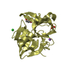
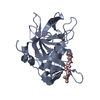
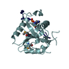
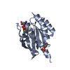
- PDB-5hma: Crystal structure of MamO protease domain from Magnetospirillum m... -
+ Open data

- Basic information
| Entry | Database: PDB / ID: 5hma | |||||||||
|---|---|---|---|---|---|---|---|---|---|---|
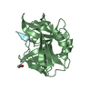
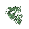
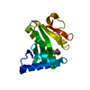
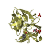
| Title | Crystal structure of MamO protease domain from Magnetospirillum magneticum (Ni bound form) | |||||||||
 Components Components |
| |||||||||
 Keywords Keywords | HYDROLASE / trypsin / biomineralization / pseudo-protease / magnetosome | |||||||||
| Function / homology |  Function and homology information Function and homology informationmagnetosome membrane / serine-type endopeptidase activity / proteolysis / metal ion binding Similarity search - Function | |||||||||
| Biological species |  Magnetospirillum magneticum (bacteria) Magnetospirillum magneticum (bacteria) | |||||||||
| Method |  X-RAY DIFFRACTION / SYNCHROTRON / MOLECULAR REPLACEMENT / Resolution: 2.299 Å X-RAY DIFFRACTION / SYNCHROTRON / MOLECULAR REPLACEMENT / Resolution: 2.299 Å | |||||||||
 Authors Authors | Hershey, D.M. / Ren, X. / Hurley, J.H. / Komeili, A. | |||||||||
| Funding support |  United States, 2items United States, 2items
| |||||||||
 Citation Citation | Journal: Plos Biol. / Year: 2016 Title: MamO Is a Repurposed Serine Protease that Promotes Magnetite Biomineralization through Direct Transition Metal Binding in Magnetotactic Bacteria. Authors: Hershey, D.M. / Ren, X. / Melnyk, R.A. / Browne, P.J. / Ozyamak, E. / Jones, S.R. / Chang, M.C. / Hurley, J.H. / Komeili, A. | |||||||||
| History |
|
- Structure visualization
| Structure viewer | Molecule: MolmilJmol/JSmol |
|---|
- Downloads & links
-Download
| PDBx/mmCIF format | 5hma.cif.gz | 88.1 KB | Display | PDBx/mmCIF format |
|---|---|---|---|---|
| PDB format | pdb5hma.ent.gz | 66.5 KB | Display | PDB format |
| PDBx/mmJSON format | 5hma.json.gz | Tree view | PDBx/mmJSON format | |
| Others |  Other downloads Other downloads |
-Validation report
| Arichive directory | https://data.pdbj.org/pub/pdb/validation_reports/hm/5hmaftp://data.pdbj.org/pub/pdb/validation_reports/hm/5hma | HTTPS FTP |
|---|
-Related structure data
| Related structure data |  5hm9C  5mh9 C: citing same article ( S: Starting model for refinement |
|---|---|
| Similar structure data |


-Links
 PDBj
PDBj
- Assembly
| Deposited unit | 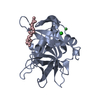
| ||||||||||||
|---|---|---|---|---|---|---|---|---|---|---|---|---|---|
| 1 |
| ||||||||||||
| Unit cell |
| ||||||||||||
| Components on special symmetry positions |
|
-Components
| #1: Protein | Mass: 19692.545 Da / Num. of mol.: 1 Source method: isolated from a genetically manipulated source Source: (gene. exp.) Magnetospirillum magneticum (strain AMB-1 / ATCC 700264) (bacteria)Strain: AMB-1 / ATCC 700264 / Gene: amb0969 / Production host: |
|---|---|
| #2: Protein/peptide | Mass: 443.539 Da / Num. of mol.: 1 Source method: isolated from a genetically manipulated source Source: (gene. exp.) Magnetospirillum magneticum (strain AMB-1 / ATCC 700264) (bacteria)Strain: AMB-1 / ATCC 700264 / Production host: |
| #3: Chemical | ChemComp-NI /   Mass: 58.693 Da / Num. of mol.: 1 / Source method: obtained synthetically / Formula: Ni Mass: 58.693 Da / Num. of mol.: 1 / Source method: obtained synthetically / Formula: Ni |
| #4: Chemical | ChemComp-CL /   Mass: 35.453 Da / Num. of mol.: 1 / Source method: obtained synthetically / Formula: Cl Mass: 35.453 Da / Num. of mol.: 1 / Source method: obtained synthetically / Formula: Cl |
| #5: Water | ChemComp-HOH /  Mass: 18.015 Da / Num. of mol.: 55 / Source method: isolated from a natural source / Formula: H2O Mass: 18.015 Da / Num. of mol.: 55 / Source method: isolated from a natural source / Formula: H2O |
| Has protein modification | N |
-Experimental details
-Experiment
| Experiment | Method: X-RAY DIFFRACTION / Number of used crystals: 1 |
|---|
- Sample preparation
| Crystal | Density Matthews: 4.47 Å3/Da / Density % sol: 72.51 % / Description: Cubic |
|---|---|
| Crystal grow | Temperature: 291 K / Method: vapor diffusion, hanging drop / pH: 4.6 / Details: 50mM Na Acetate pH 4.6, 3.6M NH4Cl, 5% glycerol / PH range: 4.0 - 5.0 |
-Data collection
| Diffraction | Mean temperature: 100 K | |||||||||||||||||||||||||||||||||||||||||||||||||||||||||||||||||||||||||||||||||||||||||||||||||||||||||||||||||||||||||||||||||||||||||||||||||||||||||||||||||||||||||||||||||||||||||||||
|---|---|---|---|---|---|---|---|---|---|---|---|---|---|---|---|---|---|---|---|---|---|---|---|---|---|---|---|---|---|---|---|---|---|---|---|---|---|---|---|---|---|---|---|---|---|---|---|---|---|---|---|---|---|---|---|---|---|---|---|---|---|---|---|---|---|---|---|---|---|---|---|---|---|---|---|---|---|---|---|---|---|---|---|---|---|---|---|---|---|---|---|---|---|---|---|---|---|---|---|---|---|---|---|---|---|---|---|---|---|---|---|---|---|---|---|---|---|---|---|---|---|---|---|---|---|---|---|---|---|---|---|---|---|---|---|---|---|---|---|---|---|---|---|---|---|---|---|---|---|---|---|---|---|---|---|---|---|---|---|---|---|---|---|---|---|---|---|---|---|---|---|---|---|---|---|---|---|---|---|---|---|---|---|---|---|---|---|---|---|---|
| Diffraction source | Source: SYNCHROTRON / Site: ALS / Beamline: 8.3.1 / Wavelength: 1.116 Å | |||||||||||||||||||||||||||||||||||||||||||||||||||||||||||||||||||||||||||||||||||||||||||||||||||||||||||||||||||||||||||||||||||||||||||||||||||||||||||||||||||||||||||||||||||||||||||||
| Detector | Type: ADSC QUANTUM 315r / Detector: CCD / Date: May 13, 2015 | |||||||||||||||||||||||||||||||||||||||||||||||||||||||||||||||||||||||||||||||||||||||||||||||||||||||||||||||||||||||||||||||||||||||||||||||||||||||||||||||||||||||||||||||||||||||||||||
| Radiation | Protocol: SINGLE WAVELENGTH / Monochromatic (M) / Laue (L): M / Scattering type: x-ray | |||||||||||||||||||||||||||||||||||||||||||||||||||||||||||||||||||||||||||||||||||||||||||||||||||||||||||||||||||||||||||||||||||||||||||||||||||||||||||||||||||||||||||||||||||||||||||||
| Radiation wavelength | Wavelength: 1.116 Å / Relative weight: 1 | |||||||||||||||||||||||||||||||||||||||||||||||||||||||||||||||||||||||||||||||||||||||||||||||||||||||||||||||||||||||||||||||||||||||||||||||||||||||||||||||||||||||||||||||||||||||||||||
| Reflection | Resolution: 2.3→50 Å / Num. obs: 16999 / % possible obs: 99.9 % / Redundancy: 12.6 % / Biso Wilson estimate: 51.17 Å2 / Rmerge(I) obs: 0.085 / Rpim(I) all: 0.025 / Rrim(I) all: 0.089 / Χ2: 1.078 / Net I/av σ(I): 29.755 / Net I/σ(I): 12.3 / Num. measured all: 214349 | |||||||||||||||||||||||||||||||||||||||||||||||||||||||||||||||||||||||||||||||||||||||||||||||||||||||||||||||||||||||||||||||||||||||||||||||||||||||||||||||||||||||||||||||||||||||||||||
| Reflection shell | Diffraction-ID: 1 / Rejects: _
|
- Processing
| Software |
| ||||||||||||||||||||||||||||||||||||||||||||||||||||||||||||||||||||||||||||||||||||||||||||||||||||
|---|---|---|---|---|---|---|---|---|---|---|---|---|---|---|---|---|---|---|---|---|---|---|---|---|---|---|---|---|---|---|---|---|---|---|---|---|---|---|---|---|---|---|---|---|---|---|---|---|---|---|---|---|---|---|---|---|---|---|---|---|---|---|---|---|---|---|---|---|---|---|---|---|---|---|---|---|---|---|---|---|---|---|---|---|---|---|---|---|---|---|---|---|---|---|---|---|---|---|---|---|---|
| Refinement | Method to determine structure: MOLECULAR REPLACEMENT Starting model: 5MH9 5mh9 Resolution: 2.299→45.666 Å / SU ML: 0.23 / Cross valid method: FREE R-VALUE / σ(F): 1.34 / Phase error: 21.78 / Stereochemistry target values: ML
| ||||||||||||||||||||||||||||||||||||||||||||||||||||||||||||||||||||||||||||||||||||||||||||||||||||
| Solvent computation | Shrinkage radii: 0.9 Å / VDW probe radii: 1.11 Å / Solvent model: FLAT BULK SOLVENT MODEL | ||||||||||||||||||||||||||||||||||||||||||||||||||||||||||||||||||||||||||||||||||||||||||||||||||||
| Displacement parameters | Biso max: 142.86 Å2 / Biso mean: 63.2227 Å2 / Biso min: 35.1 Å2 | ||||||||||||||||||||||||||||||||||||||||||||||||||||||||||||||||||||||||||||||||||||||||||||||||||||
| Refinement step | Cycle: final / Resolution: 2.299→45.666 Å
| ||||||||||||||||||||||||||||||||||||||||||||||||||||||||||||||||||||||||||||||||||||||||||||||||||||
| Refine LS restraints |
| ||||||||||||||||||||||||||||||||||||||||||||||||||||||||||||||||||||||||||||||||||||||||||||||||||||
| LS refinement shell | Refine-ID: X-RAY DIFFRACTION / Total num. of bins used: 6
| ||||||||||||||||||||||||||||||||||||||||||||||||||||||||||||||||||||||||||||||||||||||||||||||||||||
| Refinement TLS params. | Method: refined / Refine-ID: X-RAY DIFFRACTION
| ||||||||||||||||||||||||||||||||||||||||||||||||||||||||||||||||||||||||||||||||||||||||||||||||||||
| Refinement TLS group |
|
