 Movie
Movie Controller


+ Open data

- Basic information
| Entry | Database: PDB / ID: 3bh2 | ||||||
|---|---|---|---|---|---|---|---|
| Title | Structural Studies of Acetoacetate Decarboxylase | ||||||
 Components Components | Acetoacetate decarboxylase | ||||||
 Keywords Keywords | LYASE / acetoacetate decarboxylase / Plasmid / Schiff base | ||||||
| Function / homology |  Function and homology information Function and homology informationacetoacetate decarboxylase / acetoacetate decarboxylase activity / identical protein binding Similarity search - Function | ||||||
| Biological species |  Clostridium acetobutylicum ATCC 824 (bacteria) Clostridium acetobutylicum ATCC 824 (bacteria) | ||||||
| Method |  X-RAY DIFFRACTION / SYNCHROTRON / MOLECULAR REPLACEMENT / Resolution: 2.4 Å X-RAY DIFFRACTION / SYNCHROTRON / MOLECULAR REPLACEMENT / Resolution: 2.4 Å | ||||||
 Authors Authors | Ho, M. / Allen, K.N. | ||||||
 Citation Citation | Journal: Nature / Year: 2009 Title: The origin of the electrostatic perturbation in acetoacetate decarboxylase. Authors: Ho, M.C. / Menetret, J.F. / Tsuruta, H. / Allen, K.N. | ||||||
| History |
| ||||||
| Remark 999 | SEQUENCE AUTHORS STATE THAT THE MUTATION L2V HAS BEEN INTRODUCED FOR CLONING PURPOSE. |
- Structure visualization
| Structure viewer | Molecule: MolmilJmol/JSmol |
|---|
- Downloads & links
-Download
| PDBx/mmCIF format | 3bh2.cif.gz | 201 KB | Display | PDBx/mmCIF format |
|---|---|---|---|---|
| PDB format | pdb3bh2.ent.gz | 163.9 KB | Display | PDB format |
| PDBx/mmJSON format | 3bh2.json.gz | Tree view | PDBx/mmJSON format | |
| Others |  Other downloads Other downloads |
-Validation report
| Arichive directory | https://data.pdbj.org/pub/pdb/validation_reports/bh/3bh2ftp://data.pdbj.org/pub/pdb/validation_reports/bh/3bh2 | HTTPS FTP |
|---|
-Related structure data


-Links
 PDBj
PDBj- Assembly
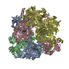
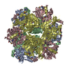
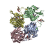
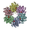
| Deposited unit | 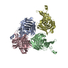
| ||||||||
|---|---|---|---|---|---|---|---|---|---|
| 1 | 
| ||||||||
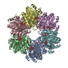
| Unit cell |
|
-Components
| #1: Protein | Mass: 27556.803 Da / Num. of mol.: 4 / Mutation: L2V Source method: isolated from a genetically manipulated source Source: (gene. exp.) Clostridium acetobutylicum ATCC 824 (bacteria)Strain: DSM 792 / JCM 1419 / LMG 5710 / VKM B-1787 / Gene: adc, CA_P0165 / Plasmid: pET3d / Production host: #2: Water | ChemComp-HOH / |  Mass: 18.015 Da / Num. of mol.: 319 / Source method: isolated from a natural source / Formula: H2O Mass: 18.015 Da / Num. of mol.: 319 / Source method: isolated from a natural source / Formula: H2O |
|---|
-Experimental details
-Experiment
| Experiment | Method: X-RAY DIFFRACTION / Number of used crystals: 1 |
|---|
- Sample preparation
| Crystal | Density Matthews: 2.73 Å3/Da / Density % sol: 54.99 % |
|---|---|
| Crystal grow | Temperature: 281 K / Method: vapor diffusion, hanging drop / pH: 5.95 Details: 40mM Phosphate buffer pH 5.95, 100mM Sarcosine, 15% PEG 3350, 18% Glycerol, VAPOR DIFFUSION, HANGING DROP, temperature 281K |
-Data collection
| Diffraction | Mean temperature: 100 K |
|---|---|
| Diffraction source | Source: SYNCHROTRON / Site: NSLS  / Beamline: X25 / Wavelength: 1 Å / Beamline: X25 / Wavelength: 1 Å |
| Detector | Type: ADSC QUANTUM 315 / Detector: CCD |
| Radiation | Protocol: SINGLE WAVELENGTH / Monochromatic (M) / Laue (L): M / Scattering type: x-ray |
| Radiation wavelength | Wavelength: 1 Å / Relative weight: 1 |
| Reflection | Resolution: 2.4→50 Å / Num. obs: 47172 / % possible obs: 98.3 % / Rmerge(I) obs: 0.093 / Net I/σ(I): 13 |
| Reflection shell | Resolution: 2.4→2.51 Å / Rmerge(I) obs: 0.354 / Mean I/σ(I) obs: 2.4 / % possible all: 93.8 |
- Processing
| Software |
| ||||||||||||||||||||||||||||||||||||||||||||||||||||||||||||||||||||||||||||||||||||||||||||||||||||||||||||||||||||||||||||||||||||||||||||||||||||||||||||||||||||||||||
|---|---|---|---|---|---|---|---|---|---|---|---|---|---|---|---|---|---|---|---|---|---|---|---|---|---|---|---|---|---|---|---|---|---|---|---|---|---|---|---|---|---|---|---|---|---|---|---|---|---|---|---|---|---|---|---|---|---|---|---|---|---|---|---|---|---|---|---|---|---|---|---|---|---|---|---|---|---|---|---|---|---|---|---|---|---|---|---|---|---|---|---|---|---|---|---|---|---|---|---|---|---|---|---|---|---|---|---|---|---|---|---|---|---|---|---|---|---|---|---|---|---|---|---|---|---|---|---|---|---|---|---|---|---|---|---|---|---|---|---|---|---|---|---|---|---|---|---|---|---|---|---|---|---|---|---|---|---|---|---|---|---|---|---|---|---|---|---|---|---|---|---|
| Refinement | Method to determine structure: MOLECULAR REPLACEMENT / Resolution: 2.4→45.79 Å / Cor.coef. Fo:Fc: 0.94 / Cor.coef. Fo:Fc free: 0.912 / SU B: 7.652 / SU ML: 0.179 / Cross valid method: THROUGHOUT / ESU R: 0.465 / ESU R Free: 0.271 / Stereochemistry target values: MAXIMUM LIKELIHOOD / Details: HYDROGENS HAVE BEEN ADDED IN THE RIDING POSITIONS
| ||||||||||||||||||||||||||||||||||||||||||||||||||||||||||||||||||||||||||||||||||||||||||||||||||||||||||||||||||||||||||||||||||||||||||||||||||||||||||||||||||||||||||
| Solvent computation | Ion probe radii: 0.8 Å / Shrinkage radii: 0.8 Å / VDW probe radii: 1.2 Å / Solvent model: MASK | ||||||||||||||||||||||||||||||||||||||||||||||||||||||||||||||||||||||||||||||||||||||||||||||||||||||||||||||||||||||||||||||||||||||||||||||||||||||||||||||||||||||||||
| Displacement parameters | Biso mean: 36.031 Å2
| ||||||||||||||||||||||||||||||||||||||||||||||||||||||||||||||||||||||||||||||||||||||||||||||||||||||||||||||||||||||||||||||||||||||||||||||||||||||||||||||||||||||||||
| Refinement step | Cycle: LAST / Resolution: 2.4→45.79 Å
| ||||||||||||||||||||||||||||||||||||||||||||||||||||||||||||||||||||||||||||||||||||||||||||||||||||||||||||||||||||||||||||||||||||||||||||||||||||||||||||||||||||||||||
| Refine LS restraints |
| ||||||||||||||||||||||||||||||||||||||||||||||||||||||||||||||||||||||||||||||||||||||||||||||||||||||||||||||||||||||||||||||||||||||||||||||||||||||||||||||||||||||||||
| LS refinement shell | Resolution: 2.4→2.46 Å / Total num. of bins used: 20
|
