 Movie
Movie Controller

[English] 日本語
 Yorodumi
- PDB-5a9e: Cryo-electron tomography and subtomogram averaging of Rous-Sarcom... -
+ Open data

- Basic information
| Entry | Database: PDB / ID: 5a9e | ||||||
|---|---|---|---|---|---|---|---|
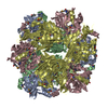
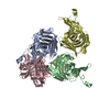
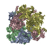
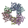
| Title | Cryo-electron tomography and subtomogram averaging of Rous-Sarcoma- Virus deltaMBD virus-like particles | ||||||
 Components Components | DELTAMBD GAG PROTEIN | ||||||
 Keywords Keywords | VIRAL PROTEIN / RETROVIRUS / ROUS-SARCOMA VIRUS / IMMATURE RETROVIRUS / VIRUS-LIKE-PARTICLE / CAPSID | ||||||
| Function / homology |  Function and homology information Function and homology informationhost cell nucleoplasm / viral procapsid maturation / host cell nucleolus / Hydrolases; Acting on peptide bonds (peptidases); Aspartic endopeptidases / viral capsid / structural constituent of virion / aspartic-type endopeptidase activity / nucleic acid binding / viral translational frameshifting / host cell plasma membrane ...host cell nucleoplasm / viral procapsid maturation / host cell nucleolus / Hydrolases; Acting on peptide bonds (peptidases); Aspartic endopeptidases / viral capsid / structural constituent of virion / aspartic-type endopeptidase activity / nucleic acid binding / viral translational frameshifting / host cell plasma membrane / proteolysis / zinc ion binding Similarity search - Function | ||||||
| Biological species |  ROUS SARCOMA VIRUS ROUS SARCOMA VIRUS | ||||||
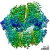
| Method | ELECTRON MICROSCOPY / electron tomography / cryo EM / Resolution: 7.7 Å | ||||||
 Authors Authors | Schur, F.K.M. / Dick, R.A. / Hagen, W.J.H. / Vogt, V.M. / Briggs, J.A.G. | ||||||
 Citation Citation | Journal: J Virol / Year: 2015 Title: The Structure of Immature Virus-Like Rous Sarcoma Virus Gag Particles Reveals a Structural Role for the p10 Domain in Assembly. Authors: Florian K M Schur / Robert A Dick / Wim J H Hagen / Volker M Vogt / John A G Briggs /   Abstract: The polyprotein Gag is the primary structural component of retroviruses. Gag consists of independently folded domains connected by flexible linkers. Interactions between the conserved capsid (CA) ...The polyprotein Gag is the primary structural component of retroviruses. Gag consists of independently folded domains connected by flexible linkers. Interactions between the conserved capsid (CA) domains of Gag mediate formation of hexameric protein lattices that drive assembly of immature virus particles. Proteolytic cleavage of Gag by the viral protease (PR) is required for maturation of retroviruses from an immature form into an infectious form. Within the assembled Gag lattices of HIV-1 and Mason-Pfizer monkey virus (M-PMV), the C-terminal domain of CA adopts similar quaternary arrangements, while the N-terminal domain of CA is packed in very different manners. Here, we have used cryo-electron tomography and subtomogram averaging to study in vitro-assembled, immature virus-like Rous sarcoma virus (RSV) Gag particles and have determined the structure of CA and the surrounding regions to a resolution of ∼8 Å. We found that the C-terminal domain of RSV CA is arranged similarly to HIV-1 and M-PMV, whereas the N-terminal domain of CA adopts a novel arrangement in which the upstream p10 domain folds back into the CA lattice. In this position the cleavage site between CA and p10 appears to be inaccessible to PR. Below CA, an extended density is consistent with the presence of a six-helix bundle formed by the spacer-peptide region. We have also assessed the affect of lattice assembly on proteolytic processing by exogenous PR. The cleavage between p10 and CA is indeed inhibited in the assembled lattice, a finding consistent with structural regulation of proteolytic maturation. IMPORTANCE: Retroviruses first assemble into immature virus particles, requiring interactions between Gag proteins that form a protein layer under the viral membrane. Subsequently, Gag is cleaved by ...IMPORTANCE: Retroviruses first assemble into immature virus particles, requiring interactions between Gag proteins that form a protein layer under the viral membrane. Subsequently, Gag is cleaved by the viral protease enzyme into separate domains, leading to rearrangement of the virus into its infectious form. It is important to understand how Gag is arranged within immature retroviruses, in order to understand how virus assembly occurs, and how maturation takes place. We used the techniques cryo-electron tomography and subtomogram averaging to obtain a detailed structural picture of the CA domains in immature assembled Rous sarcoma virus Gag particles. We found that part of Gag next to CA, called p10, folds back and interacts with CA when Gag assembles. This arrangement is different from that seen in HIV-1 and Mason-Pfizer monkey virus, illustrating further structural diversity of retroviral structures. The structure provides new information on how the virus assembles and undergoes maturation. | ||||||
| History |
|
- Structure visualization
| Movie |
Movie viewer |
|---|---|
| Structure viewer | Molecule: MolmilJmol/JSmol |
- Downloads & links
-Download
| PDBx/mmCIF format | 5a9e.cif.gz | 561.8 KB | Display | PDBx/mmCIF format |
|---|---|---|---|---|
| PDB format | pdb5a9e.ent.gz | 328.3 KB | Display | PDB format |
| PDBx/mmJSON format | 5a9e.json.gz | Tree view | PDBx/mmJSON format | |
| Others |  Other downloads Other downloads |
-Validation report
| Arichive directory | https://data.pdbj.org/pub/pdb/validation_reports/a9/5a9eftp://data.pdbj.org/pub/pdb/validation_reports/a9/5a9e | HTTPS FTP |
|---|
-Related structure data
| Related structure data | 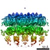 3101MC  3102C M: map data used to model this data C: citing same article ( |
|---|---|
| Similar structure data |

-Links
 PDBj
PDBj- Assembly
| Deposited unit | 
|
|---|---|
| 1 | 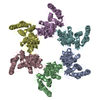
|
| 2 | 
|
| 3 | 
|
| 4 | 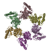
|
| 5 | 
|
| 6 | 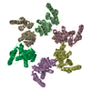
|
| 7 | 
|
-Components
| #1: Protein | Mass: 52373.262 Da / Num. of mol.: 18 Source method: isolated from a genetically manipulated source Source: (gene. exp.) ROUS SARCOMA VIRUS / Production host:  |
|---|
-Experimental details
-Experiment
| Experiment | Method: ELECTRON MICROSCOPY |
|---|---|
| EM experiment | Aggregation state: PARTICLE / 3D reconstruction method: electron tomography |
- Sample preparation
| Component | Name: IMMATURE ROUS-SARCOMA VIRUS GAG PARTICLES / Type: VIRUS |
|---|---|
| Buffer solution | Name: MES PH6.5, 100MM NACL, 2UM ZNCL2, 2MM TCEP / pH: 6.5 / Details: MES PH6.5, 100MM NACL, 2UM ZNCL2, 2MM TCEP |
| Specimen | Conc.: 5 mg/ml / Embedding applied: NO / Shadowing applied: NO / Staining applied: NO / Vitrification applied: YES |
| Specimen support | Details: HOLEY CARBON |
| Vitrification | Instrument: FEI VITROBOT MARK II / Cryogen name: ETHANE Details: DEGASSED C-FLAT 2 2-3C GRIDS WERE GLOW DISCHARGED FOR 30 SECONDS AT 20 MA. VIRUS SOLUTION WAS DILUTED IN OBS CONTAINING 10NM COLLOIDAL GOLD. 2.5UL OF THIS MIXTURE WAS APPLIED TO A GRID. ...Details: DEGASSED C-FLAT 2 2-3C GRIDS WERE GLOW DISCHARGED FOR 30 SECONDS AT 20 MA. VIRUS SOLUTION WAS DILUTED IN OBS CONTAINING 10NM COLLOIDAL GOLD. 2.5UL OF THIS MIXTURE WAS APPLIED TO A GRID. BLOTTING TIME 2.5 SECONDS. |
- Electron microscopy imaging
| Experimental equipment |  Model: Titan Krios / Image courtesy: FEI Company |
|---|---|
| Microscopy | Model: FEI TITAN KRIOS / Date: Sep 10, 2014 |
| Electron gun | Electron source:  FIELD EMISSION GUN / Accelerating voltage: 200 kV / Illumination mode: FLOOD BEAM FIELD EMISSION GUN / Accelerating voltage: 200 kV / Illumination mode: FLOOD BEAM |
| Electron lens | Mode: BRIGHT FIELD / Nominal magnification: 81000 X / Nominal defocus max: 5000 nm / Nominal defocus min: 1500 nm / Cs: 2.7 mm |
| Specimen holder | Tilt angle max: 45 ° / Tilt angle min: -45 ° |
| Image recording | Electron dose: 34 e/Å2 / Film or detector model: GATAN MULTISCAN |
| Image scans | Num. digital images: 31 |
| Radiation wavelength | Relative weight: 1 |
- Processing
| EM software |
| ||||||||||||||||||||||||
|---|---|---|---|---|---|---|---|---|---|---|---|---|---|---|---|---|---|---|---|---|---|---|---|---|---|
| CTF correction | Details: PHASE-FLIPPING OF INDIVIDUAL MICROGRAPHS | ||||||||||||||||||||||||
| Symmetry | Point symmetry: C6 (6 fold cyclic) | ||||||||||||||||||||||||
| 3D reconstruction | Method: CROSS-CORRELATION / Resolution: 7.7 Å / Num. of particles: 8375 / Nominal pixel size: 2.07 Å / Actual pixel size: 2.07 Å Details: RECONSTRUCTION CARRIED OUT USING SUBTOMOGRAM AVERAGING. SUBTOMOGRAM AVERAGING WAS PERFORMED USING SCRIPTS DERIVED FROM THE AV3 (FOERSTER, ET AL) TOM (NICKELL, ET AL) AND DYNAMO (CASTANO- ...Details: RECONSTRUCTION CARRIED OUT USING SUBTOMOGRAM AVERAGING. SUBTOMOGRAM AVERAGING WAS PERFORMED USING SCRIPTS DERIVED FROM THE AV3 (FOERSTER, ET AL) TOM (NICKELL, ET AL) AND DYNAMO (CASTANO-DIEZ, ET AL) PACKAGES. STRUCTURES FOR P10 AND CA-NTD (PDB 1P7N) AND CA-CTD (PDB 3G1I, ONE MONOMER) WERE RIGID BODY DOCKED INTO THE EM-MAP USING THE FIT-IN-MAP OPTION IN CHIMERA. REDUNDANT RESIDUES OF THE ANTIPARALLEL DIMER OF PDB 1P7N WERE REMOVED. MISSING RESIDUES IN THE HELIX 7-HELIX 8 LINKER REGION OF CA, THE CONNECTION LOOP BETWEEN P10 AND CA-NTD AND RESIDUES UPSTREAM OF THE HELIX IN P10 WERE MANUALLY MODELED USING COOT. THE FIT WAS FURTHER REFINED USING MOLECULAR DYNAMICS FLEXIBLE FITTING. SUBMISSION BASED ON EXPERIMENTAL DATA FROM EMDB EMD-3101. (DEPOSITION ID: 13603). Symmetry type: POINT | ||||||||||||||||||||||||
| Atomic model building | Protocol: FLEXIBLE FIT / Space: REAL / Details: METHOD--MOLECULAR DYNAMICS FLEXIBLE FITTING | ||||||||||||||||||||||||
| Refinement | Highest resolution: 7.7 Å | ||||||||||||||||||||||||
| Refinement step | Cycle: LAST / Highest resolution: 7.7 Å
|
