- PDB-3c8w: Crystal structure of acetoacetate decarboxylase (ADC) (YP_094708.... -
+
Open data
ID or keywords:
Loading...
-
Basic information
Entry
Database: PDB / ID: 3c8w
Title
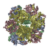
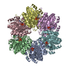
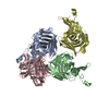
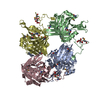
Crystal structure of acetoacetate decarboxylase (ADC) (YP_094708.1) from Legionella pneumophila subsp. pneumophila str. Philadelphia 1 at 1.60 A resolution
Components
Acetoacetate decarboxylase ADC
Keywords
LYASE / YP_094708.1 / acetoacetate decarboxylase (ADC) / Structural Genomics / Joint Center for Structural Genomics / JCSG / Protein Structure Initiative / PSI-2
Mass: 18.015 Da / Num. of mol.: 711 / Source method: isolated from a natural source / Formula: H2O
Has protein modification
Y
Sequence details
THE CONSTRUCT WAS EXPRESSED WITH A PURIFICATION TAG MGSDKIHHHHHHENLYFQG. THE TAG WAS REMOVED WITH ...THE CONSTRUCT WAS EXPRESSED WITH A PURIFICATION TAG MGSDKIHHHHHHENLYFQG. THE TAG WAS REMOVED WITH TEV PROTEASE LEAVING ONLY A GLYCINE (0) FOLLOWED BY THE TARGET SEQUENCE.
-
Experimental details
-
Experiment
Experiment
Method: X-RAY DIFFRACTION / Number of used crystals: 1
-
Sample preparation
Crystal
Density Matthews: 2.58 Å3/Da / Density % sol: 52.24 %
Crystal grow
Temperature: 277 K / Method: vapor diffusion, sitting drop / pH: 5.6 Details: NANODROP, 0.5M (NH4)2SO4, 1.0M Li2SO4, 0.1M Citrate pH 5.6, VAPOR DIFFUSION, SITTING DROP, temperature 277K
Type: MARMOSAIC 325 mm CCD / Detector: CCD / Date: Dec 10, 2007 / Details: Flat mirror (vertical focusing)
Radiation
Monochromator: Single crystal Si(111) bent (horizontal focusing) Protocol: MAD / Monochromatic (M) / Laue (L): M / Scattering type: x-ray
Radiation wavelength
ID
Wavelength (Å)
Relative weight
1
0.91837
1
2
0.97901
1
Reflection
Resolution: 1.6→29.683 Å / Num. obs: 142031 / % possible obs: 80.8 % / Observed criterion σ(I): -3 / Biso Wilson estimate: 24.188 Å2 / Rmerge(I) obs: 0.075 / Net I/σ(I): 11.27
Reflection shell
Resolution (Å)
Rmerge(I) obs
Mean I/σ(I) obs
Num. measured obs
Num. unique obs
Diffraction-ID
% possible all
1.6-1.66
0.622
1.4
37600
20973
1
66.3
1.66-1.72
0.58
1.8
48607
19568
1
71.3
1.72-1.8
0.51
2.3
80264
25102
1
80.5
1.8-1.9
0.398
3
87996
26597
1
83.2
1.9-2.02
0.273
4.5
84050
25376
1
83.6
2.02-2.17
0.193
6.3
81451
24555
1
84
2.17-2.39
0.132
9.1
85169
25700
1
84.2
2.39-2.73
0.09
13
84202
25395
1
84.6
2.73-3.44
0.046
23.4
86071
25973
1
84.9
3.44-29.683
0.023
43.1
85703
26005
1
85
-
Phasing
Phasing
Method: MAD
-
Processing
Software
Name
Version
Classification
NB
REFMAC
5.4.0067
refinement
PHENIX
refinement
SHELX
phasing
MolProbity
3beta29
modelbuilding
XSCALE
datascaling
PDB_EXTRACT
3
dataextraction
MAR345
CCD
datacollection
XDS
datareduction
SHELXD
phasing
autoSHARP
phasing
Refinement
Method to determine structure: MAD / Resolution: 1.6→29.683 Å / Cor.coef. Fo:Fc: 0.958 / Cor.coef. Fo:Fc free: 0.942 / SU B: 4.793 / SU ML: 0.079 / TLS residual ADP flag: LIKELY RESIDUAL / Cross valid method: THROUGHOUT / σ(F): 0 / ESU R: 0.094 / ESU R Free: 0.096 Stereochemistry target values: MAXIMUM LIKELIHOOD WITH PHASES Details: 1. HYDROGENS HAVE BEEN ADDED IN THE RIDING POSITIONS. 2. ATOM RECORD CONTAINS RESIDUAL B FACTORS ONLY. 3. A MET-INHIBITION PROTOCOL WAS USED FOR SELENOMETHIONINE INCORPORATION DURING PROTEIN ...Details: 1. HYDROGENS HAVE BEEN ADDED IN THE RIDING POSITIONS. 2. ATOM RECORD CONTAINS RESIDUAL B FACTORS ONLY. 3. A MET-INHIBITION PROTOCOL WAS USED FOR SELENOMETHIONINE INCORPORATION DURING PROTEIN EXPRESSION. THE OCCUPANCY OF THE SE ATOMS IN THE MSE RESIDUES WAS REDUCED TO 0.75 TO ACCOUNT FOR THE REDUCED SCATTERING POWER DUE TO PARTIAL S-MET INCORPORATION. 4. THE ELECTRON DENSITY REVEALS THAT LYSINE 125 HAS FORMED A SCHIFF BASE. IT WAS MODELED AS THE ENZYME-ACETOACETATE SCHIFF BASE INTERMEDIATE, WHICH IS CONSISTENT WITH THE OBSERVED ELECTRON DENSITY AND THE PUTATIVE ENZYMATIC MECHANISM. 5. CITRATE ANIONS FROM CRYSTALLIZATION ARE MODELED IN THIS STRUCTURE. 6. POOR ELECTRON DENSITY AROUND RESIDUE 11, 12 AND 13 IN EACH SUBUNIT RESULTS IN RAMACHANDRAN OUTLIERS.
Rfactor
Num. reflection
% reflection
Selection details
Rfree
0.228
7169
5 %
RANDOM
Rwork
0.193
-
-
-
obs
0.195
142031
93.04 %
-
Solvent computation
Ion probe radii: 0.8 Å / Shrinkage radii: 0.8 Å / VDW probe radii: 1.2 Å / Solvent model: MASK
Displacement parameters
Biso mean: 17.024 Å2
Baniso -1
Baniso -2
Baniso -3
1-
-0.06 Å2
-0.03 Å2
0 Å2
2-
-
-0.06 Å2
0 Å2
3-
-
-
0.1 Å2
Refinement step
Cycle: LAST / Resolution: 1.6→29.683 Å
Protein
Nucleic acid
Ligand
Solvent
Total
Num. atoms
7780
0
52
711
8543
Refine LS restraints
Refine-ID
Type
Dev ideal
Dev ideal target
Number
X-RAY DIFFRACTION
r_bond_refined_d
0.016
0.022
8142
X-RAY DIFFRACTION
r_bond_other_d
0.001
0.02
5459
X-RAY DIFFRACTION
r_angle_refined_deg
1.716
1.993
11130
X-RAY DIFFRACTION
r_angle_other_deg
1.044
3
13403
X-RAY DIFFRACTION
r_dihedral_angle_1_deg
5.575
5
1016
X-RAY DIFFRACTION
r_dihedral_angle_2_deg
27.738
24.076
314
X-RAY DIFFRACTION
r_dihedral_angle_3_deg
12.352
15
1271
X-RAY DIFFRACTION
r_dihedral_angle_4_deg
17.14
15
29
X-RAY DIFFRACTION
r_chiral_restr
0.097
0.2
1243
X-RAY DIFFRACTION
r_gen_planes_refined
0.007
0.021
9025
X-RAY DIFFRACTION
r_gen_planes_other
0.001
0.02
1584
X-RAY DIFFRACTION
r_mcbond_it
1.705
3
5064
X-RAY DIFFRACTION
r_mcbond_other
0.678
3
2006
X-RAY DIFFRACTION
r_mcangle_it
2.581
5
8255
X-RAY DIFFRACTION
r_scbond_it
4.337
8
3078
X-RAY DIFFRACTION
r_scangle_it
6.281
11
2875
Refine LS restraints NCS
Ens-ID: 1 / Refine-ID: X-RAY DIFFRACTION
Dom-ID
Auth asym-ID
Number
Type
Rms dev position (Å)
Weight position
1
A
1423
TIGHTPOSITIONAL
0.14
0.05
2
B
1423
TIGHTPOSITIONAL
0.12
0.05
3
C
1423
TIGHTPOSITIONAL
0.14
0.05
4
D
1423
TIGHTPOSITIONAL
0.13
0.05
1
A
1747
MEDIUMPOSITIONAL
0.21
0.5
2
B
1747
MEDIUMPOSITIONAL
0.21
0.5
3
C
1747
MEDIUMPOSITIONAL
0.38
0.5
4
D
1747
MEDIUMPOSITIONAL
0.23
0.5
1
A
1423
TIGHTTHERMAL
0.83
0.5
2
B
1423
TIGHTTHERMAL
0.88
0.5
3
C
1423
TIGHTTHERMAL
0.86
0.5
4
D
1423
TIGHTTHERMAL
0.86
0.5
1
A
1747
MEDIUMTHERMAL
0.97
2
2
B
1747
MEDIUMTHERMAL
1.03
2
3
C
1747
MEDIUMTHERMAL
1.06
2
4
D
1747
MEDIUMTHERMAL
0.99
2
LS refinement shell
Resolution: 1.6→1.642 Å / Total num. of bins used: 20
Rfactor
Num. reflection
% reflection
Rfree
0.383
437
-
Rwork
0.318
8848
-
all
-
9285
-
obs
-
-
82.89 %
Refinement TLS params.
Method: refined / Refine-ID: X-RAY DIFFRACTION
ID
L11 (°2)
L12 (°2)
L13 (°2)
L22 (°2)
L23 (°2)
L33 (°2)
S11 (Å °)
S12 (Å °)
S13 (Å °)
S21 (Å °)
S22 (Å °)
S23 (Å °)
S31 (Å °)
S32 (Å °)
S33 (Å °)
T11 (Å2)
T12 (Å2)
T13 (Å2)
T22 (Å2)
T23 (Å2)
T33 (Å2)
Origin x (Å)
Origin y (Å)
Origin z (Å)
1
0.3492
0.0683
-0.1202
0.1348
-0.1539
0.6086
0.012
0.0066
-0.0674
-0.0103
-0.0053
-0.0627
0.0726
0.0231
-0.0067
-0.0292
0.0209
0
-0.0304
-0.0112
0.0075
27.073
36.445
82.036
2
0.4066
0.1446
-0.0834
0.3742
-0.0226
0.1227
0.004
-0.0536
-0.0253
0.0457
0.0087
-0.0345
0.015
0.0323
-0.0127
-0.0114
0.0091
-0.0222
-0.0152
0.0164
-0.0264
13.083
38.735
121.155
3
0.44
-0.1896
0.0162
0.4061
-0.0264
0.1967
0.0025
0.0488
0.0208
-0.0513
0.0053
-0.0184
-0.0105
0.0277
-0.0077
-0.0194
0.0011
0.0216
-0.0081
-0.0076
-0.0329
23.984
60.018
63.726
4
0.1019
-0.0383
0.0151
0.2831
0.2363
0.353
0.0072
-0.0009
0.028
-0.0085
0.0011
-0.0809
-0.0176
0.0286
-0.0083
-0.0359
-0.0009
-0.0078
-0.0118
-0.006
0.0087
34.188
70.123
104.856
Refinement TLS group
ID
Refine-ID
Refine TLS-ID
Auth asym-ID
Label asym-ID
Auth seq-ID
Label seq-ID
1
X-RAY DIFFRACTION
1
A
A
5 - 254
6 - 255
2
X-RAY DIFFRACTION
2
B
B
6 - 254
7 - 255
3
X-RAY DIFFRACTION
3
C
C
6 - 254
7 - 255
4
X-RAY DIFFRACTION
4
D
D
5 - 254
6 - 255
+
About Yorodumi
-
News
-
Feb 9, 2022. New format data for meta-information of EMDB entries
New format data for meta-information of EMDB entries
Version 3 of the EMDB header file is now the official format.
The previous official version 1.9 will be removed from the archive.
In the structure databanks used in Yorodumi, some data are registered as the other names, "COVID-19 virus" and "2019-nCoV". Here are the details of the virus and the list of structure data.
Jan 31, 2019. EMDB accession codes are about to change! (news from PDBe EMDB page)
EMDB accession codes are about to change! (news from PDBe EMDB page)
The allocation of 4 digits for EMDB accession codes will soon come to an end. Whilst these codes will remain in use, new EMDB accession codes will include an additional digit and will expand incrementally as the available range of codes is exhausted. The current 4-digit format prefixed with “EMD-” (i.e. EMD-XXXX) will advance to a 5-digit format (i.e. EMD-XXXXX), and so on. It is currently estimated that the 4-digit codes will be depleted around Spring 2019, at which point the 5-digit format will come into force.
The EM Navigator/Yorodumi systems omit the EMD- prefix.
Related info.:Q: What is EMD? / ID/Accession-code notation in Yorodumi/EM Navigator
Yorodumi is a browser for structure data from EMDB, PDB, SASBDB, etc.
This page is also the successor to EM Navigator detail page, and also detail information page/front-end page for Omokage search.
The word "yorodu" (or yorozu) is an old Japanese word meaning "ten thousand". "mi" (miru) is to see.
Related info.:EMDB / PDB / SASBDB / Comparison of 3 databanks / Yorodumi Search / Aug 31, 2016. New EM Navigator & Yorodumi / Yorodumi Papers / Jmol/JSmol / Function and homology information / Changes in new EM Navigator and Yorodumi
 Movie
Movie Controller

 Yorodumi
 Open data
 Basic information
 Components
 Keywords
 Function and homology information
 Legionella pneumophila subsp. pneumophila str. Philadelphia 1 (bacteria)
 X-RAY DIFFRACTION /
  Authors
 Citation
 Structure visualization
 Downloads & links
 Other downloads
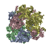

 PDBj
PDBj Assembly

 Mass: 192.124 Da / Num. of mol.: 4 / Source method: obtained synthetically / Formula: C6H8O7
 Mass: 18.015 Da / Num. of mol.: 711 / Source method: isolated from a natural source / Formula: H2O
 Sample preparation
 / Beamline: BL11-1 / Wavelength: 0.91837, 0.97901
 Processing