 Movie
Movie Controller

[English] 日本語
 Yorodumi
- PDB-3jzm: Crystal structure of the phosphorylation-site mutant T432A of the... -
+ Open data

- Basic information
| Entry | Database: PDB / ID: 3jzm | ||||||
|---|---|---|---|---|---|---|---|
| Title | Crystal structure of the phosphorylation-site mutant T432A of the KaiC circadian clock protein | ||||||
 Components Components | Circadian clock protein kinase kaiC | ||||||
 Keywords Keywords | CIRCADIAN CLOCK PROTEIN / TRANSFERASE / kaic / kinase / hexamer / ATP-binding / Biological rhythms / DNA-binding / Magnesium / Metal-binding / Nucleotide-binding / Phosphoprotein / Repressor / Serine/threonine-protein kinase / Transcription / Transcription regulation | ||||||
| Function / homology |  Function and homology information Function and homology informationregulation of phosphorelay signal transduction system / negative regulation of circadian rhythm / entrainment of circadian clock / protein serine/threonine/tyrosine kinase activity / circadian rhythm / Hydrolases; Acting on acid anhydrides; Acting on acid anhydrides to facilitate cellular and subcellular movement / non-specific serine/threonine protein kinase / protein serine kinase activity / protein serine/threonine kinase activity / regulation of DNA-templated transcription ...regulation of phosphorelay signal transduction system / negative regulation of circadian rhythm / entrainment of circadian clock / protein serine/threonine/tyrosine kinase activity / circadian rhythm / Hydrolases; Acting on acid anhydrides; Acting on acid anhydrides to facilitate cellular and subcellular movement / non-specific serine/threonine protein kinase / protein serine kinase activity / protein serine/threonine kinase activity / regulation of DNA-templated transcription / magnesium ion binding / ATP hydrolysis activity / DNA binding / ATP binding / identical protein binding Similarity search - Function | ||||||
| Biological species |  Synechococcus elongatus PCC 7942 (bacteria) Synechococcus elongatus PCC 7942 (bacteria) | ||||||
| Method |  X-RAY DIFFRACTION / SYNCHROTRON / MOLECULAR REPLACEMENT / Resolution: 2.9 Å X-RAY DIFFRACTION / SYNCHROTRON / MOLECULAR REPLACEMENT / Resolution: 2.9 Å | ||||||
 Authors Authors | Pattanayek, R. / Egli, M. / Pattanayek, S. | ||||||
 Citation Citation | Journal: Plos One / Year: 2009 Title: Structures of KaiC Circadian Clock Mutant Proteins: A New Phosphorylation Site at T426 and Mechanisms of Kinase, ATPase and Phosphatase. Authors: Pattanayek, R. / Mori, T. / Xu, Y. / Pattanayek, S. / Johnson, C.H. / Egli, M. | ||||||
| History |
|
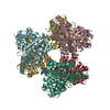
- Structure visualization
| Structure viewer | Molecule: MolmilJmol/JSmol |
|---|
- Downloads & links
-Download
| PDBx/mmCIF format | 3jzm.cif.gz | 574.3 KB | Display | PDBx/mmCIF format |
|---|---|---|---|---|
| PDB format | pdb3jzm.ent.gz | 473.8 KB | Display | PDB format |
| PDBx/mmJSON format | 3jzm.json.gz | Tree view | PDBx/mmJSON format | |
| Others |  Other downloads Other downloads |
-Validation report
| Arichive directory | https://data.pdbj.org/pub/pdb/validation_reports/jz/3jzmftp://data.pdbj.org/pub/pdb/validation_reports/jz/3jzm | HTTPS FTP |
|---|
-Related structure data
| Related structure data |  3k09C  3k0aC  3k0cC 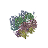 3k0eC  3k0fC  2gblS C: citing same article ( S: Starting model for refinement |
|---|---|
| Similar structure data |


-Links
 PDBj
PDBj
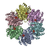
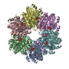
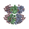
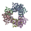
- Assembly
| Deposited unit | 
| ||||||||
|---|---|---|---|---|---|---|---|---|---|
| 1 |
| ||||||||
| Unit cell |
|
-Components
| #1: Protein | Mass: 58122.727 Da / Num. of mol.: 6 / Mutation: T432A Source method: isolated from a genetically manipulated source Source: (gene. exp.) Synechococcus elongatus PCC 7942 (bacteria)Strain: PCC7942 / Gene: kaiC, see0011, Synpcc7942_1216 / Plasmid: PET3 / Production host: References: UniProt: Q79PF4, non-specific serine/threonine protein kinase #2: Chemical | ChemComp-ATP /   Mass: 507.181 Da / Num. of mol.: 12 / Source method: obtained synthetically / Formula: C10H16N5O13P3 / Comment: ATP, energy-carrying molecule*YM Mass: 507.181 Da / Num. of mol.: 12 / Source method: obtained synthetically / Formula: C10H16N5O13P3 / Comment: ATP, energy-carrying molecule*YM#3: Chemical | ChemComp-MG /   Mass: 24.305 Da / Num. of mol.: 18 / Source method: obtained synthetically / Formula: Mg Mass: 24.305 Da / Num. of mol.: 18 / Source method: obtained synthetically / Formula: Mg#4: Water | ChemComp-HOH / |  Mass: 18.015 Da / Num. of mol.: 63 / Source method: isolated from a natural source / Formula: H2O Mass: 18.015 Da / Num. of mol.: 63 / Source method: isolated from a natural source / Formula: H2OHas protein modification | Y | |
|---|
-Experimental details
-Experiment
| Experiment | Method: X-RAY DIFFRACTION / Number of used crystals: 1 |
|---|
- Sample preparation
| Crystal | Density Matthews: 2.62 Å3/Da / Density % sol: 53.09 % |
|---|---|
| Crystal grow | Temperature: 291 K / Method: vapor diffusion, hanging drop / pH: 4 Details: SODIUM FORMATE, GLYCEROL, pH 4, VAPOR DIFFUSION, HANGING DROP, temperature 291K |
-Data collection
| Diffraction | Mean temperature: 113.15 K |
|---|---|
| Diffraction source | Source: SYNCHROTRON / Site: APS  / Beamline: 21-ID-G / Wavelength: 1 Å / Beamline: 21-ID-G / Wavelength: 1 Å |
| Detector | Type: MAR 300mm CCD / Detector: CCD / Date: Feb 24, 2008 |
| Radiation | Monochromator: Si(111) / Protocol: SINGLE WAVELENGTH / Monochromatic (M) / Laue (L): M / Scattering type: x-ray |
| Radiation wavelength | Wavelength: 1 Å / Relative weight: 1 |
| Reflection | Resolution: 2.9→6.24 Å / Num. obs: 77061 / % possible obs: 93.8 % / Rmerge(I) obs: 0.072 / Net I/σ(I): 17.8 |
| Reflection shell | Resolution: 2.9→3 Å / Rmerge(I) obs: 0.564 / Mean I/σ(I) obs: 2.8 / Num. unique all: 6624 / % possible all: 84.2 |
- Processing
| Software |
| ||||||||||||||||||||
|---|---|---|---|---|---|---|---|---|---|---|---|---|---|---|---|---|---|---|---|---|---|
| Refinement | Method to determine structure: MOLECULAR REPLACEMENT Starting model: 2GBL Resolution: 2.9→6.24 Å / σ(F): 0
| ||||||||||||||||||||
| Refinement step | Cycle: LAST / Resolution: 2.9→6.24 Å
| ||||||||||||||||||||
| LS refinement shell | Resolution: 2.9→2.92 Å
|
