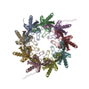
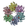
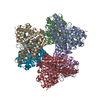
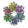
Journal: Nat Struct Mol Biol / Year: 2017 Title: Structure of the polycystic kidney disease TRP channel Polycystin-2 (PC2). Authors: Mariana Grieben / Ashley C W Pike / Chitra A Shintre / Elisa Venturi / Sam El-Ajouz / Annamaria Tessitore / Leela Shrestha / Shubhashish Mukhopadhyay / Pravin Mahajan / Rod Chalk / Nicola A ...Authors: Mariana Grieben / Ashley C W Pike / Chitra A Shintre / Elisa Venturi / Sam El-Ajouz / Annamaria Tessitore / Leela Shrestha / Shubhashish Mukhopadhyay / Pravin Mahajan / Rod Chalk / Nicola A Burgess-Brown / Rebecca Sitsapesan / Juha T Huiskonen / Elisabeth P Carpenter / Abstract: Mutations in either polycystin-1 (PC1 or PKD1) or polycystin-2 (PC2, PKD2 or TRPP1) cause autosomal-dominant polycystic kidney disease (ADPKD) through unknown mechanisms. Here we present the ...Mutations in either polycystin-1 (PC1 or PKD1) or polycystin-2 (PC2, PKD2 or TRPP1) cause autosomal-dominant polycystic kidney disease (ADPKD) through unknown mechanisms. Here we present the structure of human PC2 in a closed conformation, solved by electron cryomicroscopy at 4.2-Å resolution. The structure reveals a novel polycystin-specific 'tetragonal opening for polycystins' (TOP) domain tightly bound to the top of a classic transient receptor potential (TRP) channel structure. The TOP domain is formed from two extensions to the voltage-sensor-like domain (VSLD); it covers the channel's endoplasmic reticulum lumen or extracellular surface and encloses an upper vestibule, above the pore filter, without blocking the ion-conduction pathway. The TOP-domain fold is conserved among the polycystins, including the homologous channel-like region of PC1, and is the site of a cluster of ADPKD-associated missense variants. Extensive contacts among the TOP-domain subunits, the pore and the VSLD provide ample scope for regulation through physical and chemical stimuli.
History
Deposition
Jun 21, 2016
-
Header (metadata) release
Aug 24, 2016
-
Map release
Aug 24, 2016
-
Update
Oct 23, 2024
-
Current status
Oct 23, 2024
Processing site: PDBe / Status: Released
-
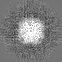
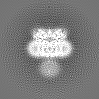
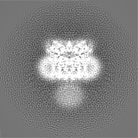
Structure visualization
Movie
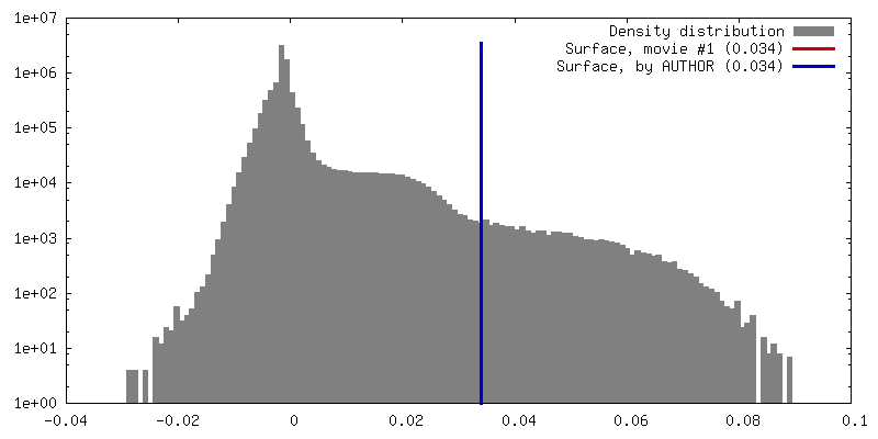
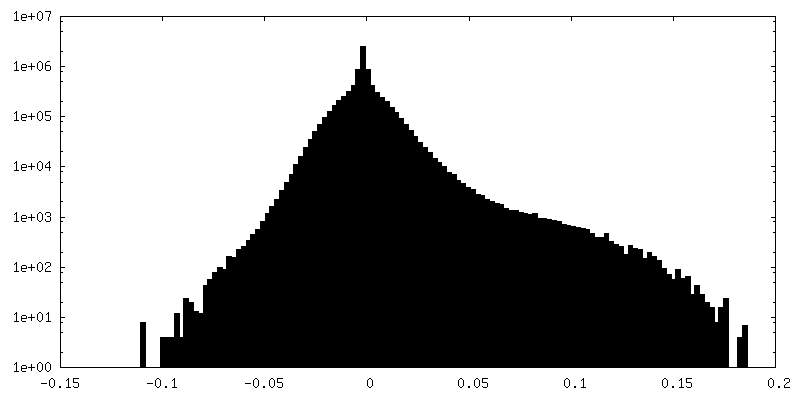
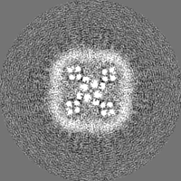
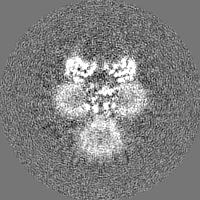
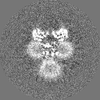
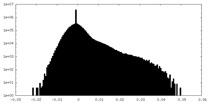
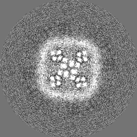
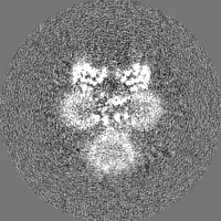
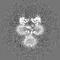
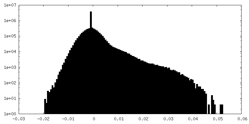
Surface view with section colored by density value
Model: C-flat / Material: COPPER / Mesh: 200 / Support film - Material: CARBON / Support film - topology: HOLEY / Pretreatment - Type: GLOW DISCHARGE / Pretreatment - Time: 15 sec. / Pretreatment - Atmosphere: AIR
Vitrification
Cryogen name: ETHANE / Chamber humidity: 90 % / Chamber temperature: 85 K / Instrument: FEI VITROBOT MARK IV Details: 3 microlitres were applied to the grid and blotted for 3secs prior to plunge in liquid ethane.
Details
Sample was monodisperse after size exclusion chromatography
-
Electron microscopy
Microscope
FEI POLARA 300
Specialist optics
Energy filter - Name: GIF Quantum / Energy filter - Lower energy threshold: 0 eV / Energy filter - Upper energy threshold: 20 eV
Image recording
Film or detector model: GATAN K2 SUMMIT (4k x 4k) / Detector mode: COUNTING / Digitization - Frames/image: 1-22 / Number grids imaged: 1 / Number real images: 758 / Average exposure time: 8.8 sec. / Average electron dose: 45.1 e/Å2 Details: Images were collected in movie-mode at 2.5 frames per second for a duration of 8.8secs
Electron beam
Acceleration voltage: 300 kV / Electron source: FIELD EMISSION GUN
Model: Tecnai Polara / Image courtesy: FEI Company
+
Image processing
Particle selection
Number selected: 161965 Details: Particles were autopicked using CTF-corrected template based picking algorithm in RELION using selected 2D class averages based on manually picked particles.
Startup model
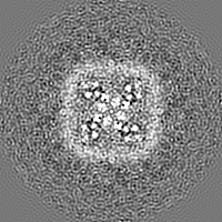
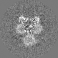
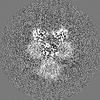
Type of model: OTHER Details: Initial model generated from a projection of a side view 2D class average
Final reconstruction
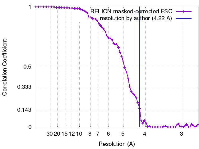
Number classes used: 1 / Applied symmetry - Point group: C4 (4 fold cyclic) / Resolution.type: BY AUTHOR / Resolution: 4.22 Å / Resolution method: FSC 0.143 CUT-OFF / Software - Name: RELION (ver. 1.3) Details: Resolution determined by gold-standard FSC procedure as implemented in RELION Number images used: 19546
In the structure databanks used in Yorodumi, some data are registered as the other names, "COVID-19 virus" and "2019-nCoV". Here are the details of the virus and the list of structure data.
Jan 31, 2019. EMDB accession codes are about to change! (news from PDBe EMDB page)
EMDB accession codes are about to change! (news from PDBe EMDB page)
The allocation of 4 digits for EMDB accession codes will soon come to an end. Whilst these codes will remain in use, new EMDB accession codes will include an additional digit and will expand incrementally as the available range of codes is exhausted. The current 4-digit format prefixed with “EMD-” (i.e. EMD-XXXX) will advance to a 5-digit format (i.e. EMD-XXXXX), and so on. It is currently estimated that the 4-digit codes will be depleted around Spring 2019, at which point the 5-digit format will come into force.
The EM Navigator/Yorodumi systems omit the EMD- prefix.
Related info.:Q: What is EMD? / ID/Accession-code notation in Yorodumi/EM Navigator
Yorodumi is a browser for structure data from EMDB, PDB, SASBDB, etc.
This page is also the successor to EM Navigator detail page, and also detail information page/front-end page for Omokage search.
The word "yorodu" (or yorozu) is an old Japanese word meaning "ten thousand". "mi" (miru) is to see.
Related info.:EMDB / PDB / SASBDB / Comparison of 3 databanks / Yorodumi Search / Aug 31, 2016. New EM Navigator & Yorodumi / Yorodumi Papers / Jmol/JSmol / Function and homology information / Changes in new EM Navigator and Yorodumi
 Movie
Movie Controller


 Open data
 Basic information
 Map data
 Sample
 Keywords
 Function and homology information
 Homo sapiens (human)
 Authors
 United Kingdom, 1 items
  Citation
 Structure visualization
 Downloads & links
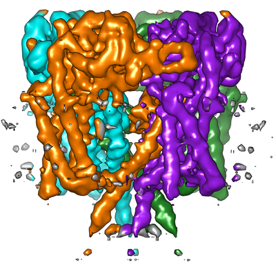 emd_8200.png
 http://ftp.pdbj.org/pub/emdb/structures/EMD-8200


 Z (Sec.)
 Y (Row.)
 X (Col.)


 Sample components
 Spodoptera frugiperda (fall armyworm)
 Processing
 Electron microscopy
 FIELD EMISSION GUN