| Entry | Database: PDB / ID: 3bgf
|
|---|
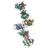
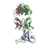
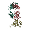
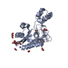
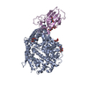
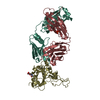
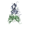
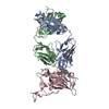
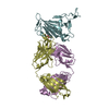
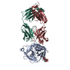
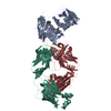
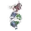
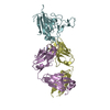
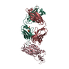
| Title | X-ray crystal structure of the SARS coronavirus spike receptor binding domain in complex with F26G19 Fab |
|---|
 Components Components | - (F26G19 Fab) x 2
- Spike protein S1
|
|---|
 Keywords Keywords | Viral Protein/Immune System / antigen-antibody complex / Envelope protein / Glycoprotein / Host-virus interaction / Membrane / Transmembrane / Virion / Virulence / Viral Protein-Immune System COMPLEX |
|---|
| Function / homology |  Function and homology information Function and homology information
Maturation of spike protein / Translation of Structural Proteins / Virion Assembly and Release / Attachment and Entry / SARS-CoV-1 activates/modulates innate immune responses / symbiont-mediated-mediated suppression of host tetherin activity / positive regulation of viral entry into host cell / membrane fusion / host cell endoplasmic reticulum-Golgi intermediate compartment membrane / receptor-mediated virion attachment to host cell ...Maturation of spike protein / Translation of Structural Proteins / Virion Assembly and Release / Attachment and Entry / SARS-CoV-1 activates/modulates innate immune responses / symbiont-mediated-mediated suppression of host tetherin activity / positive regulation of viral entry into host cell / membrane fusion / host cell endoplasmic reticulum-Golgi intermediate compartment membrane / receptor-mediated virion attachment to host cell / host cell surface receptor binding / symbiont-mediated suppression of host innate immune response / endocytosis involved in viral entry into host cell / fusion of virus membrane with host plasma membrane / fusion of virus membrane with host endosome membrane / viral envelope / host cell plasma membrane / virion membrane / membrane / identical protein bindingSimilarity search - Function Spike protein, C-terminal core receptor binding subdomain / Spike (S) protein S1 subunit, receptor-binding domain, SARS-CoV / Immunoglobulin V-set domain / Immunoglobulin C1-set domain / Spike (S) protein S1 subunit, N-terminal domain, SARS-CoV-like / Spike glycoprotein, N-terminal domain superfamily / Spike S1 subunit, receptor binding domain superfamily, betacoronavirus / Spike glycoprotein, betacoronavirus / Betacoronavirus spike (S) glycoprotein S1 subunit N-terminal (NTD) domain profile. / Spike glycoprotein S1, N-terminal domain, betacoronavirus-like ...Spike protein, C-terminal core receptor binding subdomain / Spike (S) protein S1 subunit, receptor-binding domain, SARS-CoV / Immunoglobulin V-set domain / Immunoglobulin C1-set domain / Spike (S) protein S1 subunit, N-terminal domain, SARS-CoV-like / Spike glycoprotein, N-terminal domain superfamily / Spike S1 subunit, receptor binding domain superfamily, betacoronavirus / Spike glycoprotein, betacoronavirus / Betacoronavirus spike (S) glycoprotein S1 subunit N-terminal (NTD) domain profile. / Spike glycoprotein S1, N-terminal domain, betacoronavirus-like / Betacoronavirus-like spike glycoprotein S1, N-terminal / Betacoronavirus spike (S) glycoprotein S1 subunit C-terminal (CTD) domain profile. / Spike (S) protein S1 subunit, receptor-binding domain, betacoronavirus / Betacoronavirus spike glycoprotein S1, receptor binding / Spike glycoprotein S2 superfamily, coronavirus / Spike glycoprotein S2, coronavirus, heptad repeat 1 / Spike glycoprotein S2, coronavirus, heptad repeat 2 / Coronavirus spike (S) glycoprotein S2 subunit heptad repeat 1 (HR1) region profile. / Coronavirus spike (S) glycoprotein S2 subunit heptad repeat 2 (HR2) region profile. / Spike glycoprotein S2, coronavirus / Coronavirus spike glycoprotein S2 / Alpha-Beta Plaits / Immunoglobulins / Immunoglobulin-like / Sandwich / 2-Layer Sandwich / Mainly Beta / Alpha BetaSimilarity search - Domain/homology |
|---|
| Biological species |  SARS coronavirus SARS coronavirus
  Mus musculus (house mouse) Mus musculus (house mouse) |
|---|
| Method |  X-RAY DIFFRACTION / SYNCHROTRON / MOLECULAR REPLACEMENT / molecular replacement / Resolution: 3 Å X-RAY DIFFRACTION / SYNCHROTRON / MOLECULAR REPLACEMENT / molecular replacement / Resolution: 3 Å |
|---|
 Authors Authors | Pak, J.E. / Rini, J.M. |
|---|
 Citation Citation | Journal: J.Mol.Biol. / Year: 2009
Title: Structural insights into immune recognition of the severe acute respiratory syndrome coronavirus S protein receptor binding domain.
Authors: Pak, J.E. / Sharon, C. / Satkunarajah, M. / Auperin, T.C. / Cameron, C.M. / Kelvin, D.J. / Seetharaman, J. / Cochrane, A. / Plummer, F.A. / Berry, J.D. / Rini, J.M. |
|---|
| History | | Deposition | Nov 26, 2007 | Deposition site: RCSB / Processing site: RCSB |
|---|
| Revision 1.0 | Dec 2, 2008 | Provider: repository / Type: Initial release |
|---|
| Revision 1.1 | Jul 13, 2011 | Group: Version format compliance |
|---|
| Revision 1.2 | Oct 25, 2017 | Group: Refinement description / Category: software |
|---|
| Revision 1.3 | Mar 31, 2021 | Group: Source and taxonomy / Category: entity_src_gen
Item: _entity_src_gen.pdbx_host_org_cell_line / _entity_src_gen.pdbx_host_org_strain |
|---|
| Revision 1.4 | Oct 30, 2024 | Group: Data collection / Database references ...Data collection / Database references / Refinement description / Source and taxonomy / Structure summary
Category: chem_comp_atom / chem_comp_bond ...chem_comp_atom / chem_comp_bond / database_2 / entity_src_gen / entity_src_nat / pdbx_entry_details / pdbx_initial_refinement_model / pdbx_modification_feature / struct_ncs_dom_lim / struct_ref / struct_ref_seq
Item: _database_2.pdbx_DOI / _database_2.pdbx_database_accession ..._database_2.pdbx_DOI / _database_2.pdbx_database_accession / _entity_src_gen.pdbx_beg_seq_num / _entity_src_gen.pdbx_end_seq_num / _entity_src_gen.pdbx_gene_src_gene / _entity_src_gen.pdbx_seq_type / _entity_src_nat.pdbx_beg_seq_num / _entity_src_nat.pdbx_end_seq_num / _struct_ncs_dom_lim.beg_auth_comp_id / _struct_ncs_dom_lim.end_auth_comp_id / _struct_ncs_dom_lim.end_label_asym_id / _struct_ncs_dom_lim.end_label_seq_id |
|---|
|
|---|
 Movie
Movie Controller

 Yorodumi
 Open data
 Basic information
 Structure visualization
 Downloads & links
 Other downloads
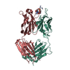


 PDBj
PDBj


 Assembly