 Movie
Movie Controller

[English] 日本語
 Yorodumi
- PDB-3b5g: Crystal Structure of the Unstable and Highly Fibrillogenic PRO7SE... -
+ Open data

- Basic information
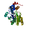
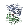
| Entry | Database: PDB / ID: 3b5g | ||||||
|---|---|---|---|---|---|---|---|
| Title | Crystal Structure of the Unstable and Highly Fibrillogenic PRO7SER Mutant of the Recombinant Variable Domain 6AJL2 | ||||||
 Components Components | AMYLOID LAMBDA 6 LIGHT CHAIN VARIABLE REGION PIP | ||||||
 Keywords Keywords | IMMUNE SYSTEM / Lambda VI subgroup / light chain variable domain / beta-sandwich / immunoglobulin / AL amyloidosis | ||||||
| Function / homology | Immunoglobulins / Immunoglobulin-like / Sandwich / Mainly Beta / ACETATE ION Function and homology information Function and homology information | ||||||
| Biological species |  Homo sapiens (human) Homo sapiens (human) | ||||||
| Method |  X-RAY DIFFRACTION / SYNCHROTRON / MOLECULAR REPLACEMENT / Resolution: 1.9 Å X-RAY DIFFRACTION / SYNCHROTRON / MOLECULAR REPLACEMENT / Resolution: 1.9 Å | ||||||
 Authors Authors | Hernandez-Santoyo, A. / Rodriguez-Romero, A. | ||||||
 Citation Citation | Journal: J.Mol.Biol. / Year: 2010 Title: A single mutation at the sheet switch region results in conformational changes favoring lambda6 light-chain fibrillogenesis. Authors: Hernandez-Santoyo, A. / del Pozo Yauner, L. / Fuentes-Silva, D. / Ortiz, E. / Rudino-Pinera, E. / Sanchez-Lopez, R. / Horjales, E. / Becerril, B. / Rodriguez-Romero, A. | ||||||
| History |
|
- Structure visualization
| Structure viewer | Molecule: MolmilJmol/JSmol |
|---|
- Downloads & links
-Download
| PDBx/mmCIF format | 3b5g.cif.gz | 61.6 KB | Display | PDBx/mmCIF format |
|---|---|---|---|---|
| PDB format | pdb3b5g.ent.gz | 46 KB | Display | PDB format |
| PDBx/mmJSON format | 3b5g.json.gz | Tree view | PDBx/mmJSON format | |
| Others |  Other downloads Other downloads |
-Validation report
| Arichive directory | https://data.pdbj.org/pub/pdb/validation_reports/b5/3b5gftp://data.pdbj.org/pub/pdb/validation_reports/b5/3b5g | HTTPS FTP |
|---|
-Related structure data
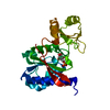
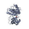
| Related structure data |  2w0kC  3bdxC  2cd0S S: Starting model for refinement C: citing same article ( |
|---|---|
| Similar structure data |


-Links
 PDBj
PDBj

- Assembly
| Deposited unit | 
| ||||||||
|---|---|---|---|---|---|---|---|---|---|
| 1 |
| ||||||||
| Unit cell |
|
-Components
| #1: Antibody | Mass: 11951.873 Da / Num. of mol.: 2 / Fragment: residues 1-111 / Mutation: P7S Source method: isolated from a genetically manipulated source Source: (gene. exp.) Homo sapiens (human) / Strain: Lambda VI light chain subgroup / Gene: VL gene segment 6a and JL2/3 gene segment / Plasmid: pSyn1 / Production host:  #2: Chemical |   Mass: 59.044 Da / Num. of mol.: 2 / Source method: obtained synthetically / Formula: C2H3O2 Mass: 59.044 Da / Num. of mol.: 2 / Source method: obtained synthetically / Formula: C2H3O2#3: Chemical | ChemComp-GOL /   Mass: 92.094 Da / Num. of mol.: 7 / Source method: obtained synthetically / Formula: C3H8O3 Mass: 92.094 Da / Num. of mol.: 7 / Source method: obtained synthetically / Formula: C3H8O3#4: Chemical | ChemComp-NA / |   Mass: 22.990 Da / Num. of mol.: 1 / Source method: obtained synthetically / Formula: Na Mass: 22.990 Da / Num. of mol.: 1 / Source method: obtained synthetically / Formula: Na#5: Water | ChemComp-HOH / |  Mass: 18.015 Da / Num. of mol.: 161 / Source method: isolated from a natural source / Formula: H2O Mass: 18.015 Da / Num. of mol.: 161 / Source method: isolated from a natural source / Formula: H2OHas protein modification | Y | Sequence details | THE RESIDUES ARE NUMBERED CONSECUTIV | |
|---|
-Experimental details
-Experiment
| Experiment | Method: X-RAY DIFFRACTION / Number of used crystals: 1 |
|---|
- Sample preparation
| Crystal | Density Matthews: 2.45 Å3/Da / Density % sol: 49.84 % |
|---|---|
| Crystal grow | Temperature: 291 K / Method: vapor diffusion, hanging drop / pH: 6.5 Details: Drops consisted of 5 microL of protein solution (7 mg/mL) plus 5 microL of Solution 7 from Crystal Screen I (Hampton Research), pH 6.5, VAPOR DIFFUSION, HANGING DROP, temperature 291K |
-Data collection
| Diffraction | Mean temperature: 103 K |
|---|---|
| Diffraction source | Source: SYNCHROTRON / Site: NSLS  / Beamline: X6A / Wavelength: 0.9791 Å / Beamline: X6A / Wavelength: 0.9791 Å |
| Detector | Type: ADSC QUANTUM 210 / Detector: CCD / Date: Sep 15, 2006 |
| Radiation | Protocol: SINGLE WAVELENGTH / Monochromatic (M) / Laue (L): M / Scattering type: x-ray |
| Radiation wavelength | Wavelength: 0.9791 Å / Relative weight: 1 |
| Reflection | Resolution: 1.9→53 Å / Num. all: 19189 / Num. obs: 19208 / % possible obs: 99.9 % / Redundancy: 11.6 % / Biso Wilson estimate: 17.9 Å2 / Rmerge(I) obs: 0.098 / Net I/σ(I): 21.4 |
| Reflection shell | Resolution: 1.9→1.949 Å / Redundancy: 9.9 % / Rmerge(I) obs: 0.36 / Mean I/σ(I) obs: 6.1 / Num. unique all: 1228 / % possible all: 99.5 |
- Processing
| Software |
| |||||||||||||||||||||||||||||||||||||||||||||||||||||||||||||||||||||||||||
|---|---|---|---|---|---|---|---|---|---|---|---|---|---|---|---|---|---|---|---|---|---|---|---|---|---|---|---|---|---|---|---|---|---|---|---|---|---|---|---|---|---|---|---|---|---|---|---|---|---|---|---|---|---|---|---|---|---|---|---|---|---|---|---|---|---|---|---|---|---|---|---|---|---|---|---|---|
| Refinement | Method to determine structure: MOLECULAR REPLACEMENT Starting model: PDB entry 2CD0 Resolution: 1.9→32.34 Å / Cor.coef. Fo:Fc: 0.959 / Cor.coef. Fo:Fc free: 0.931 / SU B: 3.648 / SU ML: 0.084 / TLS residual ADP flag: LIKELY RESIDUAL / Cross valid method: THROUGHOUT / ESU R: 0.15 / ESU R Free: 0.141 / Stereochemistry target values: MAXIMUM LIKELIHOOD / Details: HYDROGENS HAVE BEEN ADDED IN THE RIDING POSITIONS
| |||||||||||||||||||||||||||||||||||||||||||||||||||||||||||||||||||||||||||
| Solvent computation | Ion probe radii: 0.8 Å / Shrinkage radii: 0.8 Å / VDW probe radii: 1.2 Å / Solvent model: MASK | |||||||||||||||||||||||||||||||||||||||||||||||||||||||||||||||||||||||||||
| Displacement parameters | Biso mean: 19.235 Å2
| |||||||||||||||||||||||||||||||||||||||||||||||||||||||||||||||||||||||||||
| Refinement step | Cycle: LAST / Resolution: 1.9→32.34 Å
| |||||||||||||||||||||||||||||||||||||||||||||||||||||||||||||||||||||||||||
| Refine LS restraints |
| |||||||||||||||||||||||||||||||||||||||||||||||||||||||||||||||||||||||||||
| LS refinement shell | Resolution: 1.9→1.949 Å / Total num. of bins used: 20
| |||||||||||||||||||||||||||||||||||||||||||||||||||||||||||||||||||||||||||
| Refinement TLS params. | Method: refined / Refine-ID: X-RAY DIFFRACTION
| |||||||||||||||||||||||||||||||||||||||||||||||||||||||||||||||||||||||||||
| Refinement TLS group |
|
