 Movie
Movie Controller

[English] 日本語
 Yorodumi
- PDB-3b3l: Crystal structures of alternatively-spliced isoforms of human ket... -
+ Open data

- Basic information
| Entry | Database: PDB / ID: 3b3l | ||||||
|---|---|---|---|---|---|---|---|
| Title | Crystal structures of alternatively-spliced isoforms of human ketohexokinase | ||||||
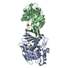
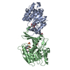
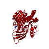
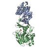
 Components Components | Ketohexokinase | ||||||
 Keywords Keywords | TRANSFERASE / Fructose kinase / Carbohydrate metabolism / Disease mutation / Phosphorylation | ||||||
| Function / homology |  Function and homology information Function and homology informationEssential fructosuria / ketohexokinase / ketohexokinase activity / regulation of glycogen metabolic process / fructose binding / Fructose catabolism / fructose metabolic process / response to insulin / protein homodimerization activity / extracellular exosome ...Essential fructosuria / ketohexokinase / ketohexokinase activity / regulation of glycogen metabolic process / fructose binding / Fructose catabolism / fructose metabolic process / response to insulin / protein homodimerization activity / extracellular exosome / ATP binding / identical protein binding / cytosol / cytoplasm Similarity search - Function | ||||||
| Biological species |  Homo sapiens (human) Homo sapiens (human) | ||||||
| Method |  X-RAY DIFFRACTION / SYNCHROTRON / MOLECULAR REPLACEMENT / Resolution: 2.9 Å X-RAY DIFFRACTION / SYNCHROTRON / MOLECULAR REPLACEMENT / Resolution: 2.9 Å | ||||||
 Authors Authors | Trinh, C.H. / Asipu, A. / Bonthron, D.T. / Phillips, S.E.V. | ||||||
 Citation Citation | Journal: To be Published Title: Crystal structures of alternatively-spliced isoforms of human ketohexokinase Authors: Trinh, C.H. / Asipu, A. / Bonthron, D.T. / Phillips, S.E.V. | ||||||
| History |
| ||||||
| Remark 999 | SEQUENCE The sequence is based on the isoform c in the database, KHK_HUMAN. |
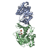
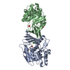
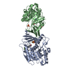
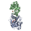
- Structure visualization
| Structure viewer | Molecule: MolmilJmol/JSmol |
|---|
- Downloads & links
-Download
| PDBx/mmCIF format | 3b3l.cif.gz | 224.6 KB | Display | PDBx/mmCIF format |
|---|---|---|---|---|
| PDB format | pdb3b3l.ent.gz | 183.3 KB | Display | PDB format |
| PDBx/mmJSON format | 3b3l.json.gz | Tree view | PDBx/mmJSON format | |
| Others |  Other downloads Other downloads |
-Validation report
| Arichive directory | https://data.pdbj.org/pub/pdb/validation_reports/b3/3b3lftp://data.pdbj.org/pub/pdb/validation_reports/b3/3b3l | HTTPS FTP |
|---|
-Related structure data
| Related structure data | 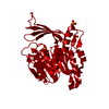 2hqqS S: Starting model for refinement |
|---|---|
| Similar structure data |


-Links
 PDBj
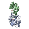
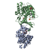
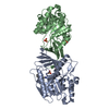
PDBj- Assembly
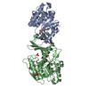
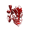
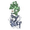
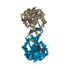
| Deposited unit | 
| ||||||||
|---|---|---|---|---|---|---|---|---|---|
| 1 | 
| ||||||||
| 2 | 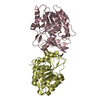
| ||||||||
| Unit cell |
|
-Components
| #1: Protein | Mass: 32561.982 Da / Num. of mol.: 4 Source method: isolated from a genetically manipulated source Source: (gene. exp.) Homo sapiens (human) / Gene: KHK / Plasmid: pET11a / Production host:  #2: Water | ChemComp-HOH / |  Mass: 18.015 Da / Num. of mol.: 18 / Source method: isolated from a natural source / Formula: H2O Mass: 18.015 Da / Num. of mol.: 18 / Source method: isolated from a natural source / Formula: H2OHas protein modification | Y | |
|---|
-Experimental details
-Experiment
| Experiment | Method: X-RAY DIFFRACTION / Number of used crystals: 1 |
|---|
- Sample preparation
| Crystal | Density Matthews: 4.38 Å3/Da / Density % sol: 71.94 % |
|---|---|
| Crystal grow | Temperature: 291 K / Method: vapor diffusion, hanging drop / pH: 4.2 Details: 20% PEG 3000, 0.1M sodium acetate, pH 4.2, VAPOR DIFFUSION, HANGING DROP, temperature 291K |
-Data collection
| Diffraction | Mean temperature: 100 K |
|---|---|
| Diffraction source | Source: SYNCHROTRON / Site: SRS  / Beamline: PX14.1 / Wavelength: 1.488 Å / Beamline: PX14.1 / Wavelength: 1.488 Å |
| Detector | Type: ADSC QUANTUM 4 / Detector: CCD / Date: Mar 13, 2004 / Details: mirrors |
| Radiation | Monochromator: Si 111 optimised for 1.488 angstrom / Protocol: SINGLE WAVELENGTH / Monochromatic (M) / Laue (L): M / Scattering type: x-ray |
| Radiation wavelength | Wavelength: 1.488 Å / Relative weight: 1 |
| Reflection | Resolution: 2.9→110.432 Å / Num. all: 49962 / Num. obs: 49962 / % possible obs: 99.7 % / Observed criterion σ(F): 0 / Observed criterion σ(I): 0 / Redundancy: 4 % / Biso Wilson estimate: 97.8 Å2 / Rmerge(I) obs: 0.06 / Rsym value: 0.06 / Net I/σ(I): 6.8 |
| Reflection shell | Resolution: 2.9→3.06 Å / Redundancy: 4 % / Rmerge(I) obs: 0.354 / Mean I/σ(I) obs: 3.6 / Num. unique all: 7208 / Rsym value: 0.354 / % possible all: 99.9 |
- Processing
| Software |
| |||||||||||||||||||||||||||||||||||||||||||||||||||||||||||||||||||||||||||||||||||||||||||||||||||||||||||||||||||||||||||||
|---|---|---|---|---|---|---|---|---|---|---|---|---|---|---|---|---|---|---|---|---|---|---|---|---|---|---|---|---|---|---|---|---|---|---|---|---|---|---|---|---|---|---|---|---|---|---|---|---|---|---|---|---|---|---|---|---|---|---|---|---|---|---|---|---|---|---|---|---|---|---|---|---|---|---|---|---|---|---|---|---|---|---|---|---|---|---|---|---|---|---|---|---|---|---|---|---|---|---|---|---|---|---|---|---|---|---|---|---|---|---|---|---|---|---|---|---|---|---|---|---|---|---|---|---|---|---|
| Refinement | Method to determine structure: MOLECULAR REPLACEMENT Starting model: PDB ENTRY 2HQQ Resolution: 2.9→25.38 Å / Cor.coef. Fo:Fc: 0.913 / Cor.coef. Fo:Fc free: 0.88 / SU B: 31.049 / SU ML: 0.276 / TLS residual ADP flag: LIKELY RESIDUAL / Isotropic thermal model: Isotropic / Cross valid method: THROUGHOUT / σ(F): 0 / ESU R: 0.577 / ESU R Free: 0.355 / Stereochemistry target values: MAXIMUM LIKELIHOOD / Details: HYDROGENS HAVE BEEN ADDED IN THE RIDING POSITIONS
| |||||||||||||||||||||||||||||||||||||||||||||||||||||||||||||||||||||||||||||||||||||||||||||||||||||||||||||||||||||||||||||
| Solvent computation | Ion probe radii: 0.8 Å / Shrinkage radii: 0.8 Å / VDW probe radii: 1.4 Å / Solvent model: MASK | |||||||||||||||||||||||||||||||||||||||||||||||||||||||||||||||||||||||||||||||||||||||||||||||||||||||||||||||||||||||||||||
| Displacement parameters | Biso mean: 62.154 Å2
| |||||||||||||||||||||||||||||||||||||||||||||||||||||||||||||||||||||||||||||||||||||||||||||||||||||||||||||||||||||||||||||
| Refinement step | Cycle: LAST / Resolution: 2.9→25.38 Å
| |||||||||||||||||||||||||||||||||||||||||||||||||||||||||||||||||||||||||||||||||||||||||||||||||||||||||||||||||||||||||||||
| Refine LS restraints |
| |||||||||||||||||||||||||||||||||||||||||||||||||||||||||||||||||||||||||||||||||||||||||||||||||||||||||||||||||||||||||||||
| LS refinement shell | Resolution: 2.9→2.975 Å / Total num. of bins used: 20
| |||||||||||||||||||||||||||||||||||||||||||||||||||||||||||||||||||||||||||||||||||||||||||||||||||||||||||||||||||||||||||||
| Refinement TLS params. | Method: refined / Refine-ID: X-RAY DIFFRACTION
| |||||||||||||||||||||||||||||||||||||||||||||||||||||||||||||||||||||||||||||||||||||||||||||||||||||||||||||||||||||||||||||
| Refinement TLS group |
|
