 Movie
Movie Controller

[English] 日本語
 Yorodumi
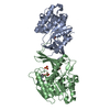
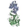
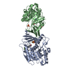
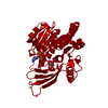
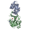
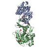
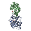
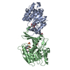
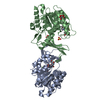
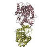
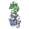
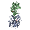
- PDB-3nbv: X-ray Structure of Ketohexokinase in complex with AMP-PNP and fructose -
+ Open data

- Basic information
| Entry | Database: PDB / ID: 3nbv | ||||||
|---|---|---|---|---|---|---|---|
| Title | X-ray Structure of Ketohexokinase in complex with AMP-PNP and fructose | ||||||
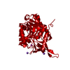
 Components Components | Ketohexokinase | ||||||
 Keywords Keywords | TRANSFERASE/TRANSFERASE INHIBITOR / ketohexokinase / TRANSFERASE / TRANSFERASE-TRANSFERASE INHIBITOR complex | ||||||
| Function / homology |  Function and homology information Function and homology informationEssential fructosuria / ketohexokinase / ketohexokinase activity / fructose binding / Fructose catabolism / regulation of glycogen metabolic process / response to sucrose / response to fructose / fructose metabolic process / response to zinc ion ...Essential fructosuria / ketohexokinase / ketohexokinase activity / fructose binding / Fructose catabolism / regulation of glycogen metabolic process / response to sucrose / response to fructose / fructose metabolic process / response to zinc ion / response to glucose / response to insulin / protein homodimerization activity / extracellular exosome / ATP binding / identical protein binding / cytoplasm / cytosol Similarity search - Function | ||||||
| Biological species |  Homo sapiens (human) Homo sapiens (human) | ||||||
| Method |  X-RAY DIFFRACTION / SYNCHROTRON / MOLECULAR REPLACEMENT / Resolution: 2.3 Å X-RAY DIFFRACTION / SYNCHROTRON / MOLECULAR REPLACEMENT / Resolution: 2.3 Å | ||||||
 Authors Authors | Abad, M.C. / Gibbs, A.C. | ||||||
 Citation Citation | Journal: J.Med.Chem. / Year: 2010 Title: Electron density guided fragment-based lead discovery of ketohexokinase inhibitors. Authors: Gibbs, A.C. / Abad, M.C. / Zhang, X. / Tounge, B.A. / Lewandowski, F.A. / Struble, G.T. / Sun, W. / Sui, Z. / Kuo, L.C. | ||||||
| History |
|
- Structure visualization
| Structure viewer | Molecule: MolmilJmol/JSmol |
|---|
- Downloads & links
-Download
| PDBx/mmCIF format | 3nbv.cif.gz | 131 KB | Display | PDBx/mmCIF format |
|---|---|---|---|---|
| PDB format | pdb3nbv.ent.gz | 100.4 KB | Display | PDB format |
| PDBx/mmJSON format | 3nbv.json.gz | Tree view | PDBx/mmJSON format | |
| Others |  Other downloads Other downloads |
-Validation report
| Arichive directory | https://data.pdbj.org/pub/pdb/validation_reports/nb/3nbvftp://data.pdbj.org/pub/pdb/validation_reports/nb/3nbv | HTTPS FTP |
|---|
-Related structure data
| Related structure data | 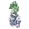 3nbwC 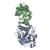 3nc2C 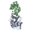 3nc9C 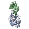 3ncaC  2hlzS C: citing same article ( S: Starting model for refinement |
|---|---|
| Similar structure data |


-Links
 PDBj
PDBj


- Assembly
| Deposited unit | 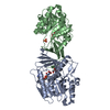
| ||||||||
|---|---|---|---|---|---|---|---|---|---|
| 1 |
| ||||||||
| Unit cell |
|
-Components
| #1: Protein | Mass: 34076.586 Da / Num. of mol.: 2 Source method: isolated from a genetically manipulated source Source: (gene. exp.) Homo sapiens (human) / Gene: KHK / Plasmid: pET28s / Production host:  References: UniProt: P50053-2, UniProt: P50053*PLUS, ketohexokinase #2: Chemical |   Mass: 506.196 Da / Num. of mol.: 2 / Source method: obtained synthetically / Formula: C10H17N6O12P3 / Comment: AMP-PNP, energy-carrying molecule analogue*YM Mass: 506.196 Da / Num. of mol.: 2 / Source method: obtained synthetically / Formula: C10H17N6O12P3 / Comment: AMP-PNP, energy-carrying molecule analogue*YM#3: Sugar | ChemComp-FRU / | 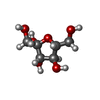  Type: D-saccharide, beta linking / Mass: 180.156 Da / Num. of mol.: 1 Type: D-saccharide, beta linking / Mass: 180.156 Da / Num. of mol.: 1Source method: isolated from a genetically manipulated source Formula: C6H12O6 #4: Chemical | ChemComp-SO4 / |   Mass: 96.063 Da / Num. of mol.: 1 / Source method: obtained synthetically / Formula: SO4 Mass: 96.063 Da / Num. of mol.: 1 / Source method: obtained synthetically / Formula: SO4#5: Water | ChemComp-HOH / |  Mass: 18.015 Da / Num. of mol.: 120 / Source method: isolated from a natural source / Formula: H2O Mass: 18.015 Da / Num. of mol.: 120 / Source method: isolated from a natural source / Formula: H2O |
|---|
-Experimental details
-Experiment
| Experiment | Method: X-RAY DIFFRACTION / Number of used crystals: 1 |
|---|
- Sample preparation
| Crystal | Density Matthews: 3.58 Å3/Da / Density % sol: 65.65 % |
|---|---|
| Crystal grow | Temperature: 295 K / Method: vapor diffusion, sitting drop / pH: 4.5 Details: 17% PEG 8k, 0.1M Na-Citrate, 0.2M Ammonium sulfate, pH 4.5, VAPOR DIFFUSION, SITTING DROP, temperature 295K |
-Data collection
| Diffraction | Mean temperature: 200 K |
|---|---|
| Diffraction source | Source: SYNCHROTRON / Site: APS  / Beamline: 17-ID / Wavelength: 1 Å / Beamline: 17-ID / Wavelength: 1 Å |
| Detector | Type: ADSC QUANTUM 210 / Detector: CCD / Date: Apr 5, 2007 / Details: mirror |
| Radiation | Monochromator: double crystal mono, Si(111) from Accel / Protocol: SINGLE WAVELENGTH / Monochromatic (M) / Laue (L): M / Scattering type: x-ray |
| Radiation wavelength | Wavelength: 1 Å / Relative weight: 1 |
| Reflection | Resolution: 2.2→50 Å / Num. obs: 49441 / Observed criterion σ(F): 0 / Observed criterion σ(I): -3 / Redundancy: 4.7 % / Rmerge(I) obs: 0.071 / Rsym value: 0.066 / Net I/σ(I): 18.9 |
| Reflection shell | Resolution: 2.2→2.28 Å / Redundancy: 4.6 % / Mean I/σ(I) obs: 2.85 / Num. unique all: 4843 / % possible all: 97.8 |
- Processing
| Software |
| ||||||||||||||||||||||||||||||||||||||||||||||||||||||||||||||||||||||||||||||||||||||||||||||||||
|---|---|---|---|---|---|---|---|---|---|---|---|---|---|---|---|---|---|---|---|---|---|---|---|---|---|---|---|---|---|---|---|---|---|---|---|---|---|---|---|---|---|---|---|---|---|---|---|---|---|---|---|---|---|---|---|---|---|---|---|---|---|---|---|---|---|---|---|---|---|---|---|---|---|---|---|---|---|---|---|---|---|---|---|---|---|---|---|---|---|---|---|---|---|---|---|---|---|---|---|
| Refinement | Method to determine structure: MOLECULAR REPLACEMENT Starting model: PDB ENTRY 2HLZ Resolution: 2.3→41.041 Å / SU ML: 0.32 / Isotropic thermal model: anisotropic / σ(F): 1.34 / σ(I): 0 / Stereochemistry target values: ML
| ||||||||||||||||||||||||||||||||||||||||||||||||||||||||||||||||||||||||||||||||||||||||||||||||||
| Solvent computation | Shrinkage radii: 0.9 Å / VDW probe radii: 1.11 Å / Solvent model: FLAT BULK SOLVENT MODEL / Bsol: 62.236 Å2 / ksol: 0.4 e/Å3 | ||||||||||||||||||||||||||||||||||||||||||||||||||||||||||||||||||||||||||||||||||||||||||||||||||
| Displacement parameters |
| ||||||||||||||||||||||||||||||||||||||||||||||||||||||||||||||||||||||||||||||||||||||||||||||||||
| Refinement step | Cycle: LAST / Resolution: 2.3→41.041 Å
| ||||||||||||||||||||||||||||||||||||||||||||||||||||||||||||||||||||||||||||||||||||||||||||||||||
| Refine LS restraints |
| ||||||||||||||||||||||||||||||||||||||||||||||||||||||||||||||||||||||||||||||||||||||||||||||||||
| LS refinement shell |
|
