glycolytic process through fructose-1-phosphate / Fructose catabolism / : / fructose catabolic process / ketohexokinase / ketohexokinase activity / regulation of glycogen metabolic process / regulation of glycogen biosynthetic process / fructose binding / response to sucrose ...glycolytic process through fructose-1-phosphate / Fructose catabolism / : / fructose catabolic process / ketohexokinase / ketohexokinase activity / regulation of glycogen metabolic process / regulation of glycogen biosynthetic process / fructose binding / response to sucrose / fructose metabolic process / carbohydrate catabolic process / response to zinc ion / response to glucose / response to insulin / protein homodimerization activity / ATP binding / identical protein binding / nucleus / cytosol / cytoplasm Similarity search - Function
Mass: 18.015 Da / Num. of mol.: 389 / Source method: isolated from a natural source / Formula: H2O
Has ligand of interest
Y
Has protein modification
Y
-
Experimental details
-
Experiment
Experiment
Method: X-RAY DIFFRACTION / Number of used crystals: 1
-
Sample preparation
Crystal
Density Matthews: 1.79 Å3/Da / Density % sol: 31.32 %
Crystal grow
Temperature: 290.15 K / Method: vapor diffusion, hanging drop / pH: 7 Details: 1.2 M Ammonium Citrate Tribasic, 20% glycerol, 1.3 M KNO3, 100 mM MgCl2, 220 mM fructose, and 52 mM ADP
-
Data collection
Diffraction
Mean temperature: 100 K / Serial crystal experiment: N
Diffraction source
Source: ROTATING ANODE / Type: BRUKER AXS MICROSTAR / Wavelength: 1.54 Å
Detector
Type: Bruker Platinum 135 / Detector: CCD / Date: Dec 22, 2016
Radiation
Protocol: SINGLE WAVELENGTH / Monochromatic (M) / Laue (L): M / Scattering type: x-ray
Radiation wavelength
Wavelength: 1.54 Å / Relative weight: 1
Reflection
Resolution: 1.79→37.96 Å / Num. obs: 23632 / % possible obs: 97 % / Redundancy: 1.88 % / Biso Wilson estimate: 9.33 Å2 / CC1/2: 0.99 / Rmerge(I) obs: 0.14 / Rrim(I) all: 0.15 / Net I/σ(I): 14.3
Reflection shell
Resolution: 1.79→1.86 Å / Rmerge(I) obs: 0.3 / Mean I/σ(I) obs: 6.4 / Num. unique obs: 2058 / CC1/2: 0.95 / % possible all: 74
-
Processing
Software
Name
Version
Classification
PHENIX
1.13_2998
refinement
PHENIX
1.13_2998
refinement
SADABS
datascaling
PDB_EXTRACT
3.25
dataextraction
SAINT
datareduction
PHENIX
1.10.1_2155
phasing
Refinement
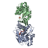
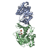
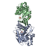
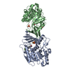
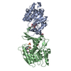
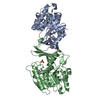
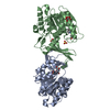
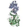
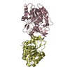
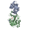
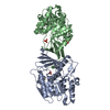
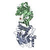
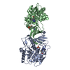
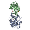
Method to determine structure: MOLECULAR REPLACEMENT Starting model: 3Q92
In the structure databanks used in Yorodumi, some data are registered as the other names, "COVID-19 virus" and "2019-nCoV". Here are the details of the virus and the list of structure data.
Jan 31, 2019. EMDB accession codes are about to change! (news from PDBe EMDB page)
EMDB accession codes are about to change! (news from PDBe EMDB page)
The allocation of 4 digits for EMDB accession codes will soon come to an end. Whilst these codes will remain in use, new EMDB accession codes will include an additional digit and will expand incrementally as the available range of codes is exhausted. The current 4-digit format prefixed with “EMD-” (i.e. EMD-XXXX) will advance to a 5-digit format (i.e. EMD-XXXXX), and so on. It is currently estimated that the 4-digit codes will be depleted around Spring 2019, at which point the 5-digit format will come into force.
The EM Navigator/Yorodumi systems omit the EMD- prefix.
Related info.:Q: What is EMD? / ID/Accession-code notation in Yorodumi/EM Navigator
Yorodumi is a browser for structure data from EMDB, PDB, SASBDB, etc.
This page is also the successor to EM Navigator detail page, and also detail information page/front-end page for Omokage search.
The word "yorodu" (or yorozu) is an old Japanese word meaning "ten thousand". "mi" (miru) is to see.
Related info.:EMDB / PDB / SASBDB / Comparison of 3 databanks / Yorodumi Search / Aug 31, 2016. New EM Navigator & Yorodumi / Yorodumi Papers / Jmol/JSmol / Function and homology information / Changes in new EM Navigator and Yorodumi
 Movie
Movie Controller

 Yorodumi
 Open data
 Basic information
 Components
 Keywords
 Function and homology information
 X-RAY DIFFRACTION /
  Authors
 Citation
 Structure visualization
 Downloads & links
 Other downloads
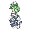


 PDBj
PDBj

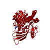
 Assembly
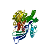


 Mass: 427.201 Da / Num. of mol.: 1
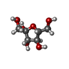
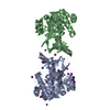
 Type: D-saccharide, beta linking / Mass: 180.156 Da / Num. of mol.: 1 / Source method: obtained synthetically / Formula: C6H12O6 / Feature type: SUBJECT OF INVESTIGATION
 Mass: 62.005 Da / Num. of mol.: 1 / Source method: obtained synthetically / Formula: NO3
 Mass: 18.015 Da / Num. of mol.: 389 / Source method: isolated from a natural source / Formula: H2O
 Sample preparation
 Processing