 Movie
Movie Controller

[English] 日本語
 Yorodumi
- PDB-6ul7: Structure of human ketohexokinase-C in complex with fructose, NO3... -
+ Open data

- Basic information
| Entry | Database: PDB / ID: 6ul7 | ||||||
|---|---|---|---|---|---|---|---|
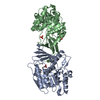
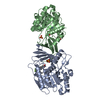
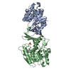
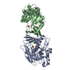
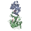
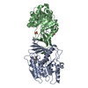
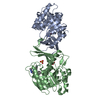
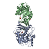
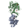
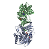
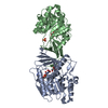
| Title | Structure of human ketohexokinase-C in complex with fructose, NO3, and osthole | ||||||
 Components Components | Ketohexokinase | ||||||
 Keywords Keywords | TRANSFERASE / beta-clasp / sugar kinase / PfKB family / KHK / ketohexokinase / fructose / osthole | ||||||
| Function / homology |  Function and homology information Function and homology informationEssential fructosuria / ketohexokinase / ketohexokinase activity / fructose binding / Fructose catabolism / regulation of glycogen metabolic process / response to sucrose / response to fructose / fructose metabolic process / response to zinc ion ...Essential fructosuria / ketohexokinase / ketohexokinase activity / fructose binding / Fructose catabolism / regulation of glycogen metabolic process / response to sucrose / response to fructose / fructose metabolic process / response to zinc ion / response to glucose / response to insulin / protein homodimerization activity / extracellular exosome / ATP binding / identical protein binding / cytoplasm / cytosol Similarity search - Function | ||||||
| Biological species |  Homo sapiens (human) Homo sapiens (human) | ||||||
| Method |  X-RAY DIFFRACTION / MOLECULAR REPLACEMENT / Resolution: 2.3 Å X-RAY DIFFRACTION / MOLECULAR REPLACEMENT / Resolution: 2.3 Å | ||||||
 Authors Authors | Gasper, W.C. / Gardner, S. / Allen, K.N. / Tolan, D.R. | ||||||
 Citation Citation | Journal: To be Published Title: Structure of human ketohexokinase-C in complex with fructose, NO3, and osthole Authors: Gasper, W.C. / Gardner, S. / Ross, A. / Allen, K.N. / Tolan, D.R. | ||||||
| History |
|
- Structure visualization
| Structure viewer | Molecule: MolmilJmol/JSmol |
|---|
- Downloads & links
-Download
| PDBx/mmCIF format | 6ul7.cif.gz | 78.5 KB | Display | PDBx/mmCIF format |
|---|---|---|---|---|
| PDB format | pdb6ul7.ent.gz | 55.1 KB | Display | PDB format |
| PDBx/mmJSON format | 6ul7.json.gz | Tree view | PDBx/mmJSON format | |
| Others |  Other downloads Other downloads |
-Validation report
| Arichive directory | https://data.pdbj.org/pub/pdb/validation_reports/ul/6ul7ftp://data.pdbj.org/pub/pdb/validation_reports/ul/6ul7 | HTTPS FTP |
|---|
-Related structure data
| Related structure data | 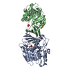 3q92S S: Starting model for refinement |
|---|---|
| Similar structure data |


-Links
 PDBj
PDBj

- Assembly
| Deposited unit | 
| ||||||||||||
|---|---|---|---|---|---|---|---|---|---|---|---|---|---|
| 1 | 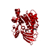
| ||||||||||||
| Unit cell |
| ||||||||||||
| Components on special symmetry positions |
|
-Components
| #1: Protein | Mass: 34733.336 Da / Num. of mol.: 1 Source method: isolated from a genetically manipulated source Source: (gene. exp.) Homo sapiens (human) / Gene: KHK / Plasmid: pET28a / Production host:  |
|---|---|
| #2: Sugar | ChemComp-FRU /   Type: D-saccharide, beta linking / Mass: 180.156 Da / Num. of mol.: 1 Type: D-saccharide, beta linking / Mass: 180.156 Da / Num. of mol.: 1Source method: isolated from a genetically manipulated source Formula: C6H12O6 |
| #3: Chemical | ChemComp-A0O /   Mass: 244.286 Da / Num. of mol.: 1 / Source method: isolated from a natural source / Formula: C15H16O3 / Feature type: SUBJECT OF INVESTIGATION Mass: 244.286 Da / Num. of mol.: 1 / Source method: isolated from a natural source / Formula: C15H16O3 / Feature type: SUBJECT OF INVESTIGATION |
| #4: Chemical | ChemComp-NO3 /   Mass: 62.005 Da / Num. of mol.: 1 / Source method: isolated from a natural source / Formula: NO3 Mass: 62.005 Da / Num. of mol.: 1 / Source method: isolated from a natural source / Formula: NO3 |
| #5: Water | ChemComp-HOH /  Mass: 18.015 Da / Num. of mol.: 199 / Source method: isolated from a natural source / Formula: H2O Mass: 18.015 Da / Num. of mol.: 199 / Source method: isolated from a natural source / Formula: H2O |
| Has ligand of interest | Y |
-Experimental details
-Experiment
| Experiment | Method: X-RAY DIFFRACTION / Number of used crystals: 1 |
|---|
- Sample preparation
| Crystal | Density Matthews: 3.37 Å3/Da / Density % sol: 63.5 % |
|---|---|
| Crystal grow | Temperature: 290.15 K / Method: vapor diffusion, hanging drop / pH: 5.5 Details: 0.1 M Bis-Tris, pH 5.5, 2 M ammonium sulfate, 1.3 M potassium nitrate, 100 mM magnesium chloride, 220 mM fructose, 3.8 mM osthole |
-Data collection
| Diffraction | Mean temperature: 100 K / Serial crystal experiment: N |
|---|---|
| Diffraction source | Source: ROTATING ANODE / Type: BRUKER AXS MICROSTAR / Wavelength: 1.54 Å |
| Detector | Type: Bruker Platinum 135 / Detector: CCD / Date: Jan 12, 2018 |
| Radiation | Protocol: SINGLE WAVELENGTH / Monochromatic (M) / Laue (L): M / Scattering type: x-ray |
| Radiation wavelength | Wavelength: 1.54 Å / Relative weight: 1 |
| Reflection | Resolution: 2.3→47.79 Å / Num. obs: 20332 / % possible obs: 97 % / Redundancy: 46.4 % / Biso Wilson estimate: 33.25 Å2 / CC1/2: 0.97 / Net I/σ(I): 18.5 |
| Reflection shell | Resolution: 2.3→2.38 Å / Mean I/σ(I) obs: 4.5 / Num. unique obs: 1568 / CC1/2: 0.83 / % possible all: 76 |
- Processing
| Software |
| ||||||||||||||||||
|---|---|---|---|---|---|---|---|---|---|---|---|---|---|---|---|---|---|---|---|
| Refinement | Method to determine structure: MOLECULAR REPLACEMENT Starting model: PDB entry 3Q92 Resolution: 2.3→47.79 Å / Cross valid method: THROUGHOUT
| ||||||||||||||||||
| Displacement parameters | Biso max: 78.55 Å2 / Biso mean: 33.252 Å2 / Biso min: 12.32 Å2 | ||||||||||||||||||
| Refinement step | Cycle: LAST / Resolution: 2.3→47.79 Å
| ||||||||||||||||||
| LS refinement shell | Resolution: 2.3001→2.3823 Å
|
